Breast cancer image information bottleneck multi-task classification and segmentation method and system

An image information and multi-task technology, applied in the field of medical image processing, can solve problems such as no theoretical understanding of the internal organizational structure, controversial task-related interpretability, and lack of solutions, so as to improve interpretability, improve accuracy, Model Robust Effects

Active Publication Date: 2021-07-30
SHANDONG NORMAL UNIV
View PDF8 Cites 2 Cited by
  • Summary
  • Abstract
  • Description
  • Claims
  • Application Information

AI Technical Summary

Problems solved by technology

[0004]Current multi-task collaborative learning methods have achieved remarkable results in many fields, but the interpretability of task associations has been a controversial issue
Despite their widespread application in recent years, deep learning models for multi-tasking are still black-bo

Method used


Image

  • Breast cancer image information bottleneck multi-task classification and segmentation method and system
  • Breast cancer image information bottleneck multi-task classification and segmentation method and system
  • Breast cancer image information bottleneck multi-task classification and segmentation method and system

Examples


Embodiment 1

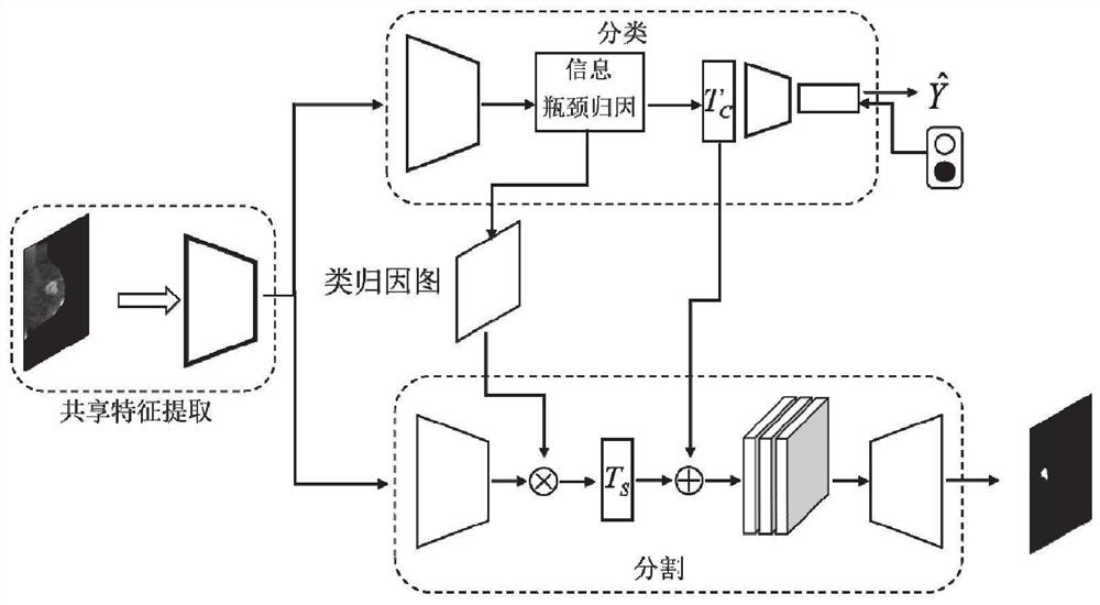
[0042] Such as figure 1 As shown, the breast cancer image information bottleneck multi-task classification and segmentation method of the present embodiment includes:

[0043] S101: Obtain several breast images of contrast-enhanced X-ray photography and corresponding benign and malignant categories and pixel-level annotations of tumor positions.

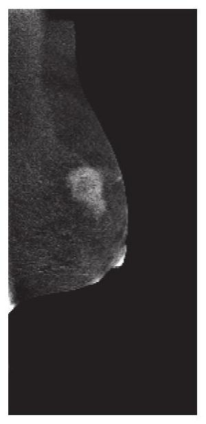
[0044] In a specific implementation, several contrast-enhanced X-ray mammography images are obtained, such as Figure 2(a)-Figure 2(d) As shown, including the CC and MLO positions of each patient's left and right breasts.

[0045] S102: Perform a preprocessing operation on each acquired contrast-enhanced X-ray mammography image.

[0046] Specifically, the preprocessing operations include cropping, random image enhancement, image normalization, and scale adjustment.

[0047] In the cropping process, each acquired breast image is cropped into an image block of 512*512 pixels to obtain a cropped breast energy spectrum image, such as ...

Embodiment 2

[0098] Such as Figure 4 As shown, this embodiment provides a breast cancer image information bottleneck multi-task classification and segmentation system, which includes:

[0099] A data acquisition module, which is configured to: acquire a plurality of breast images of contrast-enhanced X-ray photography and corresponding benign and malignant categories and pixel-level annotations of tumor positions;

[0100] A data preprocessing module, which is configured to: perform a preprocessing operation on each breast image obtained from contrast-enhanced X-ray photography;

[0101] A shared feature extraction module, which is configured to: use a multi-task network to extract a multi-task shared representation from each breast image after preprocessing;

[0102] The breast benign and malignant classification module is configured to: input the shared representation to the classification encoder to obtain the encoding tensor of the intermediate layer, and then send the intermediate e...

Embodiment 3

[0106] This embodiment provides a computer-readable storage medium, on which a computer program is stored, and when the program is executed by a processor, the steps in the above-mentioned method for breast cancer image information bottleneck multi-task classification and segmentation are realized.


PUM


Abstract

The invention belongs to the technical field of medical image processing, and provides a breast cancer image information bottleneck multi-task classification and segmentation method and system. The method comprises the following steps: acquiring a plurality of breast images of contrast enhanced X-ray photography and corresponding benign and malignant categories and lump position pixel-level labels; performing preprocessing operation on each acquired breast image of the contrast enhanced X-ray photography; adopting a multi-task network to extract multi-task shared representation for each preprocessed mammary gland image; inputting the shared representation to a classification encoder to obtain an encoding tensor of an intermediate layer, and then sending the intermediate encoding tensor to an information bottleneck attribution module for feature compression and benign and malignant classification to obtain a classification task tensor; adapting the shared representation to a segmentation network to obtain a segmentation task tensor; and carrying out feature fusion on the classification task tensor and the segmentation task tensor to segment the focus.

Description

technical field [0001] The invention belongs to the technical field of medical image processing, and in particular relates to a breast cancer image information bottleneck multi-task classification and segmentation method and system. Background technique [0002] The statements in this section merely provide background information related to the present invention and do not necessarily constitute prior art. [0003] Early detection and diagnosis of breast cancer is the key to improving survival and efficacy. Regular physical exams and screenings can help detect breast lumps and suspicious lesions early. Mammography, a commonly used method of breast cancer screening, contributes significantly to reducing breast cancer mortality through early detection of cancer. Breast cancer can be identified based on the position of the radiologist and the analysis of lesions in the images, but this results in a diagnosis that is subject to subjective factors and clinical diagnostic variab...

Claims


Application Information

Patent Timeline
IPC IPC(8): G06K9/62G06T7/11G06T7/00G06N3/04G06N3/08
CPCG06T7/11G06T7/0012G06N3/084G06T2207/10116G06T2207/20081G06T2207/20084G06T2207/20172G06T2207/20221G06T2207/30068G06T2207/30096G06N3/045G06F18/2415G06F18/253G06F18/214
Inventor 郑元杰王军霞宋景琦马骏姜岩芸
Owner SHANDONG NORMAL UNIV
Why Eureka
Try Eureka