All-optical X-ray microscopic imaging system

A microscopic imaging and X-ray technology, applied in the direction of using wave/particle radiation, material analysis using radiation, instruments, etc., can solve the problems of sample projection image overlap, complex optical path adjustment system, and difficult optical path adjustment, etc., so as to facilitate popularization Easy to use, simple system structure, simple image reconstruction

Pending Publication Date: 2021-08-03
北京市辐射中心
View PDF14 Cites 1 Cited by
  • Summary
  • Abstract
  • Description
  • Claims
  • Application Information

AI Technical Summary

Problems solved by technology

[0004] However, since the micropores of the porous shutter 3 cannot be aligned with the micropores of the rear polycapillary X-ray lens 4, the projection images of the subsequent samples will overlap.
This not only reduces the efficiency of the imaging system, but also complicates the image reconstruction process
[0005] Moreover, the existing system has many optical devices in its beam splitter, so it is difficult to adjust the optical path, and requires a complex optical path adjustment system, which is difficult to be widely promoted

Method used


Image

  • All-optical X-ray microscopic imaging system
  • All-optical X-ray microscopic imaging system
  • All-optical X-ray microscopic imaging system

Examples


Embodiment

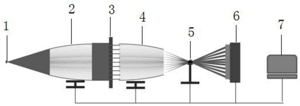
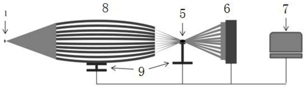
[0043] X-ray generator 1 can be combined with laboratory point focal spot X-ray tube and beam splitter 8, the focus of the light tube is placed at the front focal spot of the beam splitter, and the beam splitter 8 scatters the received X-ray tube The X-ray beams are converted into respective independent narrow cone beams, and the cone beams are converged at the rear focal point of the beam splitter 8 . The X-ray generator 1 is a rotating anode molybdenum target light source, the focal spot diameter is 300 microns, and the power is 3 kilowatts. The inner diameter of the sub-tube of the beam splitter 8 is 1 micron, the outer diameter is 30 microns, the working distance of the beam splitter 8 is 15 mm, and the focal spot size 30 microns. The sample 5 is adjusted and placed at the focal spot of the beam splitter 8 through the precision displacement platform 9 , and the imaging detector 5 is placed behind it, and the projection images of the sample 3 at various angles can be obtain...


PUM


Abstract

The invention provides an all-optical X-ray microscopic imaging system which comprises an X-ray generator and further comprises a beam splitter, wherein the beam splitter is used for splitting an X-ray beam emitted by the X-ray generator into independent conical X-ray light beams and converging the X-ray light beams at the focal point behind the X-ray light beams, and the converging position is the position where a focal spot is located; a sample is placed at the focal spot; the beam splitter is manufactured in a manufacturing mode of manufacturing a multi-capillary X-ray lens; adjacent sub-tubes of the beam splitter are formed by drawing thick-wall glass tubes; and the diameter D of the sub-tube is greater than the size of the focal spot. The beam splitter adopted by the system provided by the invention has enough thickness, so that light beams emitted from the beam splitter can be separated, a finally formed projection image has no ghosting, and the imaging efficiency of the system is high; and the system is simple in structure, low in cost and convenient to popularize and use.

Description

technical field [0001] The invention relates to the technical field of optical imaging, in particular to an all-optical X-ray microscopic imaging system. Background technique [0002] X-ray imaging technology with high time and space resolution is of great significance to chemistry, physics, biology, energy, new materials, environment and other related research fields, and is a hot and difficult point in X-ray imaging research. The current technology used to obtain three-dimensional imaging of samples is mainly CT imaging technology or limited-angle CT imaging technology, but it is necessary to obtain the projection of the sample from one angle to another, and it is difficult to obtain projections from different angles of the sample in one exposure. In 2020, the Plenoptic X-ray microscope (Plenoptic X-raymicroscopy) proposed by P. Korecki's team combined with the characteristics of a plenoptic camera and a polycapillary X-ray lens can achieve one exposure to obtain one hundr...

Claims


Application Information

Patent Timeline
Patent Type & Authority Applications(China)
IPC IPC(8): G01N23/04G01B15/04
CPCG01N23/04G01B15/04
Inventor 孙学鹏张晓芸孙天希李成波
Owner 北京市辐射中心
Why Eureka
Try Eureka