Time-resolved fluorescence immunochromatography method for simultaneously detecting aflatoxin B1 and zearalenone toxin in corn

A time-resolved fluorescence, aflatoxin technology, applied in the field of detection

Pending Publication Date: 2021-10-22
HEILONGJIANG PONY TESTING TECH CO LTD
View PDF0 Cites 1 Cited by
  • Summary
  • Abstract
  • Description
  • Claims
  • Application Information

AI Technical Summary

Problems solved by technology

[0005] However, time-resolved fluorescent microspheres are rarely reported as immunochromatographic detection methods for the simultaneous detection of multiple mycotoxins as immunoprobes.

Method used


Image

  • Time-resolved fluorescence immunochromatography method for simultaneously detecting aflatoxin B1 and zearalenone toxin in corn

Examples


Embodiment 1

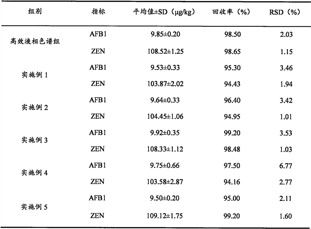
[0017] (1) The method of using carboxylated fluorescent microspheres to prepare fluorescent probes is as follows: 10 μL of time-resolved fluorescent microspheres are evenly dispersed in 1 mL of activation solution, and 50 μL of EDC solution (0.5 mg / mL) and 50 μL of NHS solution (0.5 mg / mL) are sequentially added. / mL), ultrasonically mixed, placed on a shaker (200r / min) to activate at room temperature for 10min, centrifuged (14000r / min, 20min) to discard the supernatant; Resuspend; add antibody, place on a shaking table and shake at room temperature for 20 minutes; then add 20 μL of blocking solution, place on a shaking table and shake at room temperature for blocking reaction for 1 hour, centrifuge to discard the supernatant; reconstitute with 200 μL of fluorescent microsphere reconstitution solution, and store at 4°C. Spray (0.8μL / cm) coated antigen AFB1-OVA and coated antigen ZEN-OVA on the NC membrane as the detection line (T1 line and T2 line), goat anti-mouse IgG as the q...

Embodiment 2

[0021] (1) The method of using carboxylated fluorescent microspheres to prepare fluorescent probes is as follows: 10 μL of time-resolved fluorescent microspheres are evenly dispersed in 1 mL of activation solution, and 50 μL of EDC solution (0.5 mg / mL) and 50 μL of NHS solution (0.5 mg / mL) are sequentially added. / mL), ultrasonically mixed, placed on a shaker (200r / min) to activate at room temperature for 12min, centrifuged (14000r / min, 20min) to discard the supernatant; Resuspend; add antibody, place on shaker and shake at room temperature for 25 minutes; then add 20 μL of blocking solution, place on shaker and shake at room temperature for blocking reaction for 1 hour, centrifuge to discard supernatant; reconstitute with 200 μL of fluorescent microsphere reconstitution solution, and store at 4°C. Spray (0.8μL / cm) coated antigen AFB1-OVA and coated antigen ZEN-OVA on the NC membrane as the detection line (T1 line and T2 line), goat anti-mouse IgG as the quality control line (C...

Embodiment 3

[0026] (1) The method of using carboxylated fluorescent microspheres to prepare fluorescent probes is as follows: 10 μL of time-resolved fluorescent microspheres are evenly dispersed in 1 mL of activation solution, and 50 μL of EDC solution (0.5 mg / mL) and 50 μL of NHS solution (0.5 mg / mL) are sequentially added. / mL), ultrasonically mixed, placed on a shaker (200r / min) to activate at room temperature for 15min, centrifuged (14000r / min, 20min) to discard the supernatant; Resuspend; add antibody, place on a shaker and shake at room temperature for 30 minutes; then add 20 μL of blocking solution, place on a shaker and shake at room temperature for blocking reaction for 1 hour, centrifuge to discard the supernatant; reconstitute with 200 μL of fluorescent microsphere reconstitution solution, and store at 4°C. Spray (0.8μL / cm) coated antigen AFB1-OVA and coated antigen ZEN-OVA on the NC membrane as the detection line (T1 line and T2 line), goat anti-mouse IgG as the quality control...


PUM


Abstract

The invention discloses a time-resolved fluorescence immunochromatography method for simultaneously detecting aflatoxin B1 and zearalenone toxin in corn. The method comprises the following steps: (1) preparing a time-resolved fluorescence immunochromatography test strip; (2) screening detection conditions of the time-resolved fluorescence immunochromatography test strip; and (3) detecting the sample, wherein the detection mainly comprises the following steps: (3-1) pre-treating the sample; (3-2) screening an extraction solvent and extraction time; (3-3) detection and analysis: adding100 mu L of the to-be-detected sample solution into the micropores containing the fluorescent probe, uniformly mixing the solution, and incubating the solution at 37 DEG C for 2-5 minutes, and inserting the test strip into the micropore, reacting at 37 DEG C for 5-10 minutes, and then quantitatively / qualitatively evaluating the judgment result of the fluorescence chromatography test strip. The time-resolved fluorescence immunochromatography detection method established by the invention has the advantages of simultaneous detection of aflatoxin B1 and ochratoxin A, high accuracy, short detection time, simple operation and the like.

Description

technical field [0001] The invention belongs to the technical field of detection, and mainly relates to a time-resolved fluorescent immunochromatographic method for simultaneously detecting aflatoxin B1 and erythralenone toxin in corn. Background technique [0002] Aflatoxin B1 (AFB1) is the most carcinogenic of all mycotoxins, and is classified as a class I carcinogen by the International Agency for Research on Cancer (IARC). It can cause acute hepatitis, and long-term intake can even cause liver cancer. In order to ensure people's food safety, the National Quality Supervision Bureau requires AFB1 to be a mandatory inspection item for cereal foods. The national standard GB 2761-2017 "Mycotoxin Limits in Food" stipulates that the maximum residue limit of AFB1 in cereals and their products is 5.0-20μg / kg. [0003] Zearalenone (ZEN), also known as F2 toxin, is a non-steroidal mycotoxin mainly produced by Fusarium graminearum and Fusarium roseum. One of the most widely cont...

Claims


Application Information

Patent Timeline
Patent Type & Authority Applications(China)
IPC IPC(8): G01N33/577G01N33/558G01N33/543G01N33/533G01N33/53G01N21/64
CPCG01N33/577G01N33/54313G01N33/558G01N33/533G01N33/5308G01N21/6408G01N21/6428
Inventor 王中江郭增旺赫志强韩丽杰王卓
Owner HEILONGJIANG PONY TESTING TECH CO LTD
Why Eureka
Try Eureka