Composite cone beam CT enhanced contrast agent and preparation method thereof

A configuration method and cone beam technology, applied in the field of oral and maxillofacial medical imaging, can solve problems such as troubles, and achieve the effect of improving the development rate and depth of development

Active Publication Date: 2021-11-02
NANJING STOMATOLOGICAL HOSPITAL
View PDF8 Cites 0 Cited by
  • Summary
  • Abstract
  • Description
  • Claims
  • Application Information

AI Technical Summary

Problems solved by technology

[0003] The diagnosis of cracked teeth has important clinical significance for the design of reasonable treatment plan and the judgment of prognosis. However, due to the small and hidden cracks in cracked teeth, the variety of clinical manifestations in different clinical stages, and overlapping with many dental diseases, The diagnosis of cracked teeth is a clinically troubled problem
In recent years, oral cone-beam CT has made it possible to diagnose cracked teeth due to its extremely high spatial resolution. There is still room for improvement in the accuracy, sensitivity and specificity of CBCT in the diagnosis of cracked teeth

Method used


Image

  • Composite cone beam CT enhanced contrast agent and preparation method thereof
  • Composite cone beam CT enhanced contrast agent and preparation method thereof
  • Composite cone beam CT enhanced contrast agent and preparation method thereof

Examples


Embodiment 1

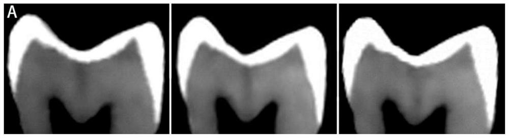
[0025] Such as figure 1 and figure 2 As shown, a composite cone beam CT enhanced contrast agent, including: X-ray imaging agent sodium iodide, distilled water, hydrophilic pharmaceutical penetrant dimethyl sulfoxide, organic solvent and bacteriolytic agent ethanol, lipophilic Penetrant and flavoring agent ethyl acetate.

[0026] A compound cone-beam CT enhanced contrast agent configuration method, comprising the following steps:

[0027] First, slowly dissolve 25 grams of 99% dimethyl sulfoxide in 10 g of distilled water, stir and dissolve to a homogeneous solution, then add 10 grams of 99% ethanol, stir and dissolve to a homogeneous solution, then add 99% ethyl acetate 10 g, stirred to a homogeneous solution; finally, 30 g of 99% sodium iodide was added to dissolve to a homogeneous solution to obtain a composite cone-beam CT enhanced contrast agent that can be used in the diagnosis of cracked teeth in vivo.

[0028] In order to clearly understand the contrast effect of th...

Embodiment 2

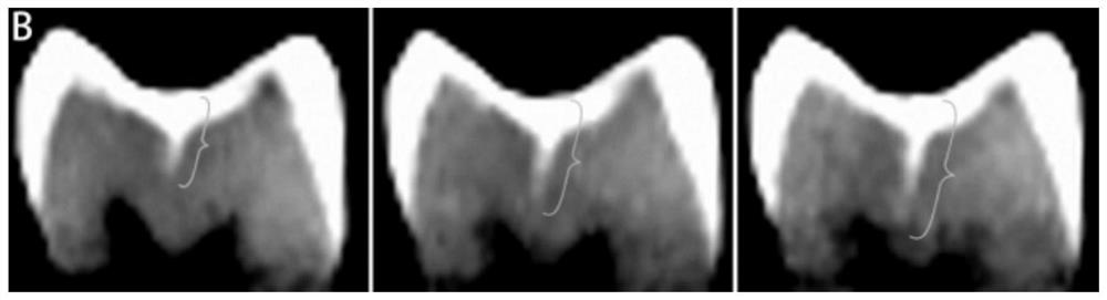
[0031] Such as image 3 As shown, first slowly dissolve 25 grams of 99% dimethyl sulfoxide in 10 g of distilled water, stir and dissolve to a homogeneous solution, then add 10 grams of 99% ethanol, stir and dissolve to a homogeneous solution, and finally add 99% 10 grams of ethyl acetate, stirred to a homogeneous solution; finally a composite CT enhanced contrast agent that can be used for the diagnosis of cracked teeth in vivo is obtained.

[0032] Use a soft-bristled brush to pick up the composite CT-enhanced contrast agent, apply it on the surface of the cracked tooth extracted in vivo, and wait for 1 hour. After the contrast agent penetrates into the crack, perform a CBCT scan to determine whether the crown is present. Cracks and crack depths.

Embodiment 3

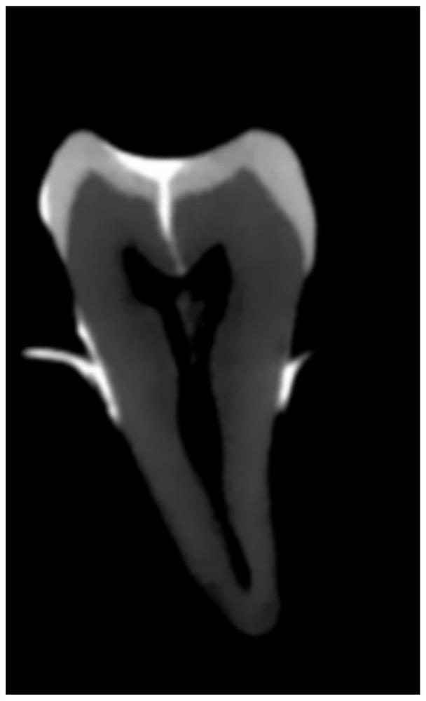
[0034] Such as Figure 4-Figure 6 As shown, first 35 grams of 99% sodium iodide are dissolved in 25 grams of 99% dimethyl sulfoxide, stirred and dissolved to a homogeneous solution, then 10 grams of 99% ethanol is added, stirred and dissolved to a homogeneous solution, Finally, 10 grams of 99% ethyl acetate was added and stirred until a homogeneous solution was obtained; finally, a composite CT enhanced contrast agent that can be used for in vivo cracked tooth diagnosis was obtained.

[0035] Collect the longitudinally fractured teeth extracted in vivo, remove the filling in the pulp cavity, use a dropper with a capacity of 1.8ml to absorb the contrast medium, and drop the contrast medium into the pulp cavity, wait for 15 minutes, and wait for the contrast medium to penetrate After entering the crack, a CBCT scan is performed to determine whether there is a crack in the tooth root.

[0036] Depend on figure 1 It can be seen that the hidden cracks under the body condition are...


PUM


Abstract

The invention relates to the technical field of oral and maxillofacial medical imaging technologies, in particular to a composite cone beam CT enhanced contrast agent which comprises an X-ray imaging agent sodium iodide, distilled water, a hydrophilic medicinal penetrant dimethyl sulfoxide, an organic solvent, a fungicide ethanol, a lipophilic penetrant and a flavoring agent ethyl acetate. The imaging component content of the contrast agent can be adjusted, and the contrast agent can be recognized with different densities of tooth tissues and various fillers, so that the accuracy, sensitivity and specificity of CBCT on tooth subfissure diagnosis are further improved; a preparation method of a composite cone beam CT enhanced contrast agent comprises the following steps: firstly, slowly dissolving 25 g of 99% dimethyl sulfoxide in 10 g of distilled water, stirring and dissolving to obtain a homogeneous solution, then adding 10 g of 99% ethanol, stirring and dissolving to obtain a homogeneous solution, then adding 10 g of 99% ethyl acetate, stirring to obtain a homogeneous solution, and finally adding 10 g of 99% ethyl acetate, stirring to obtain a homogeneous solution; finally, 30 g of 99% sodium iodide is added and dissolved into a homogeneous solution, and compared with meglumine diatrizoate, the contrast agent prepared through the method has the advantages that the developing rate and the developing depth are obviously improved.

Description

technical field [0001] The invention relates to the technical field of oral and maxillofacial medical imaging, in particular to a compound cone-beam CT enhanced contrast agent and a configuration method thereof. Background technique [0002] Cracked teeth are one of the three main causes of tooth loss in adults following periodontal disease and dental caries. At present, cracked teeth in the broad sense are divided into 5 categories: enamel surface cracks, cusp fractures, cracked teeth, longitudinally fractured teeth, and vertical root fractures. Facing the fracture line extending in the direction of the apex, the fourth type of cracked tooth refers to the fractured tooth that continues from the crown to the root, and the fifth type of VRF refers to the tooth with a fractured root. [0003] The diagnosis of cracked teeth has important clinical significance for the design of reasonable treatment plan and the judgment of prognosis. However, due to the small and hidden cracks ...

Claims


Application Information

Patent Timeline
Patent Type & Authority Applications(China)
IPC IPC(8): A61K49/04
CPCA61K49/0438Y02A50/30
Inventor 林梓桐胡子洋王铁梅高安天曹丹彤觧鑫
Owner NANJING STOMATOLOGICAL HOSPITAL
Why Eureka
Try Eureka