Cervix uteri abnormal cell recognition method and device and electronic equipment

A technology of abnormal cells and cervical cells, applied in the field of image recognition, can solve the problems of restricting the recognition speed of the system, not fully considering the principle of P16 staining of cervical cells, and unable to effectively obtain the staining intensity of abnormal cells, so as to improve the speed of the algorithm and the recognition of effect of effect

Pending Publication Date: 2022-01-28
北京智源人工智能研究院
View PDF0 Cites 1 Cited by
  • Summary
  • Abstract
  • Description
  • Claims
  • Application Information

AI Technical Summary

Problems solved by technology

However, the methods in the prior art have the following deficiencies for P16-stained cervical cells: First, all image processing operations are based on the RGB color space, without fully considering the principle of cervical cell P16 staining, and the positive expression of abnormal cells is forcibly compared with the RGB value. Correlation cannot effectively obtain the staining intensity of abnormal cells; moreover, try to extract and classify each detected cell, which greatly restricts the recognition speed of the system
Unable to meet clinical requirements for data processing speed and real-time performance

Method used


Image

  • Cervix uteri abnormal cell recognition method and device and electronic equipment
  • Cervix uteri abnormal cell recognition method and device and electronic equipment
  • Cervix uteri abnormal cell recognition method and device and electronic equipment

Examples


Embodiment 1

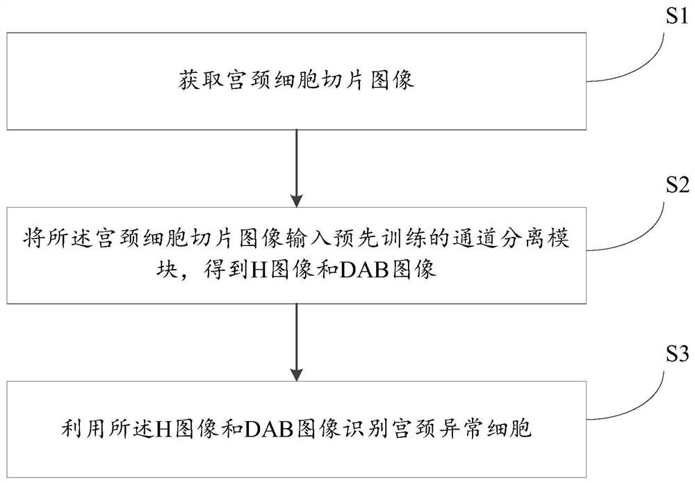
[0062] like figure 1 As shown, the embodiment of the present invention firstly provides a method for identifying abnormal cervical cells, which is characterized in that it includes:

[0063] S1. Acquiring images of cervical cell slices;

[0064] S2. Input the cervical cell slice image into the pre-trained channel separation module to obtain an H image and a DAB image, the H image is a hematoxylin stained image, and the DAB image is an immunohistochemical stained image;

[0065] S3. Using the H image and the DAB image to identify abnormal cells of the cervix.

[0066] The above-mentioned abnormal cervical cell identification method effectively separates the P16 stained image in RGB space into hematoxylin stained image H and immunohistochemical stained image DAB (diaminobenzidine stained image) through an adaptive cervical cell P16 stained image channel separation algorithm. ), where the H channel image mainly shows nuclear staining, and the DAB channel image mainly shows the ...

Embodiment 2

[0129] like Figure 5 As shown, another aspect of the present invention also includes a functional module architecture that is completely consistent with the aforementioned method flow, that is, an embodiment of the present invention also provides a device for identifying abnormal cervical cells, including:

[0130] An acquisition module 201, configured to acquire cervical cell slice images;

[0131] A calculation module 202, configured to input the cervical cell slice image into a pre-trained channel separation module to obtain an H image and a DAB image, the H image is a hematoxylin stained image, and the DAB image is an immunohistochemical stained image;

[0132] An identification module 203, configured to identify abnormal cells of the cervix by using the H image and the DAB image

[0133] The device can be realized by the method for identifying abnormal cervical cells provided in the first embodiment above, and the specific implementation method can refer to the descript...


PUM


Abstract

The invention discloses a cervix uteri abnormal cell recognition method and device and electronic equipment. The method comprises the steps that a cervix uteri cell section image is acquired; the cervical cell slice image is input into a pre-trained channel separation module to obtain an H image and a DAB image, with the H image being a hematoxylin stained image, and the DAB image being an immunohistochemical stained image; and abnormal cervical cells are identified by using the H image and the DAB image. According to the technical scheme, the cell detection speed can be obviously increased in cervical cancer positive cell detection, the recognition accuracy is enhanced, and the algorithm execution efficiency is improved.

Description

technical field [0001] The invention relates to the technical field of image recognition, in particular to a method, device and electronic equipment for identifying abnormal cervical cells. [0002] This invention is the research result of the major project of Science and Technology Innovation 2030 "Innovative Development and Application of Artificial Intelligence Pilot Area" (2020AAA0105203). Background technique [0003] Cervical cancer is an important cause of female-specific cancer-related death. If patients are diagnosed at the precancerous stage or earlier, the cure rate can be as high as 98%, and the mortality rate will be significantly reduced. In recent years, medical microscopic image processing technology using computer image processing, artificial intelligence and other technologies is developing rapidly. At present, the identification of abnormal cells in cervical cancer is generally carried out by the following methods: in the image RGB space, the location of ...

Claims


Application Information

Patent Timeline
Patent Type & Authority Applications(China)
IPC IPC(8): G06V20/69G06V10/82G06T7/00G06T7/11G06T7/194G06N3/08
CPCG06T7/0012G06T7/11G06T7/194G06N3/088G06T2207/10024G06T2207/20032G06T2207/20081G06T2207/20084G06T2207/10056
Inventor 赵帅袁莎曹岗赵健
Owner 北京智源人工智能研究院
Why Eureka
Try Eureka