Method for inducing muscle stem cell expansion

A technology of muscle cells and stem cells, applied in the material field of inducing the expansion of adult muscle stem cells, which can solve the problems of insufficient donor cells, loss of damage repair, loss of stemness of muscle stem cells, etc.

Pending Publication Date: 2022-02-22
CENT FOR EXCELLENCE IN MOLECULAR CELL SCI CHINESE ACAD OF SCI
View PDF6 Cites 0 Cited by
  • Summary
  • Abstract
  • Description
  • Claims
  • Application Information

AI Technical Summary

Problems solved by technology

The biggest difficulty in muscle stem cell therapy is the shortage of donor cells
Muscle stem cells cultured

Method used


Image

  • Method for inducing muscle stem cell expansion
  • Method for inducing muscle stem cell expansion
  • Method for inducing muscle stem cell expansion

Examples


Embodiment 1

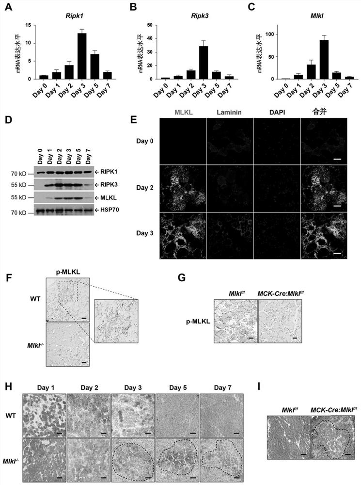
[0235] Example 1. After acute muscle injury, RIPK1-RIPK3-MLKL is highly expressed in muscle fiber cells and programmed necrosis occurs.

[0236] In the process of muscle degeneration after muscle injury, myofiber cells undergo necrosis (Chargé and Rudnicki, 2004). We established an acute injury model of tibialis anterior muscle by injecting cardiotoxin (CTX), so as to study whether necroptosis is involved in the repair process of muscle tissue injury. CTX is a polypeptide isolated from snake venom, which can cause severe contraction of muscle fiber cells by excessively activating nerves, resulting in depolarization of muscle fiber cells and damage to cell membranes, resulting in muscle damage (Chargé and Rudnicki, 2004; Shiau et al. ,1976). It is widely used in the study of skeletal muscle regeneration (Garry et al., 2016; Hardy et al., 2016).

[0237] The modeling method was to inject a total of 100 μl of CTX solution with a concentration of 10 μM into the tibialis anterior...

Embodiment 2

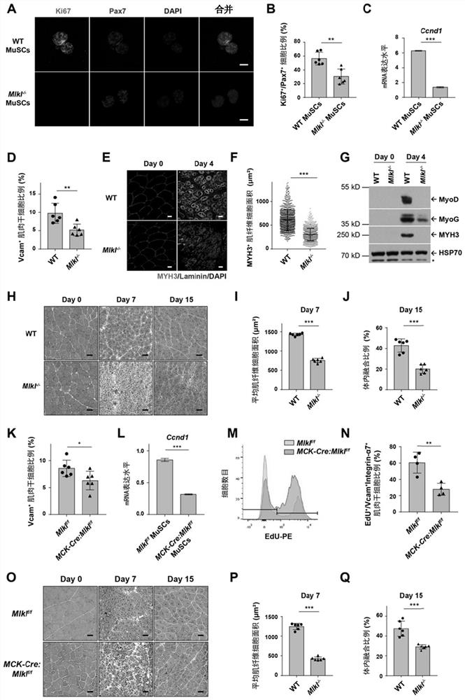
[0241] Example 2. The absence of programmed necrosis in muscle fiber cells affects the proliferation of muscle stem cells and the process of muscle injury repair.

[0242] In order to further investigate the effect of the blockage of myofibroblast necrosis, we compared the wild-type and Mlkl after muscle injury by FACS analysis. - / - Differences in the proportion of mouse muscle stem cells. On the 3rd day after muscle injury, the muscle stem cells were selected for immunofluorescence staining, and the results were shown in Mlkl - / - Ki67 in mouse + Pax7 + The proportion of cells also decreased ( figure 2 , A-B). Real-time quantitative PCR showed that the cell cycle protein Cyclin D1 in Mlkl - / - Compared with wild-type mice, the mRNA level in mouse muscle stem cells was significantly lower, indicating that the degree of expansion of muscle stem cells was significantly different from that of wild-type mice ( figure 2 , C-D), implying that Mlkl - / - Muscle stem cells prolif...

Embodiment 3

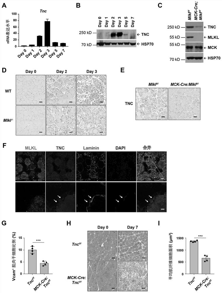
[0245] Example 3, muscle fiber cells with programmed necrosis highly express TNC and release it outside the cells to promote the proliferation of muscle stem cells.

[0246] Through a series of in-depth studies, the inventors found that muscle fiber cells undergoing programmed necrosis can promote the rapid proliferation of muscle stem cells after injury by releasing tenascin C. There is almost no mRNA expression of Tnc in normal TA. The results of real-time quantitative PCR and immunoblotting showed that the mRNA and protein levels of Tnc in TA were significantly increased after CTX injury ( image 3 , A-B). Consistent with it, the results of TNC immunohistochemical staining after TA injury showed that there was no expression of TNC in normal muscle fiber cells, but the expression of TNC was significantly increased in the damaged muscle fiber cells with swelling morphology after injury in wild-type mice ( image 3, D, upper row). After 3 days of CTX injury in wild-type mic...


PUM


Abstract

The invention relates to programmed necrosis of muscle fiber cells after muscle tissue injury and a proliferation mechanism of muscle stem cells and application of the programmed necrosis and the proliferation mechanism. Muscle fibers with programmed necrosis highly express and release TNC, and the TNC promotes rapid proliferation of muscle stem cells by combining and activating EGFR on the surfaces of the muscle stem cells, so that damaged muscle tissues are repaired. The invention also provides a composition containing TNC, which can promote in-vitro mass amplification and long-term passage of muscle stem cells.

Description

technical field [0001] The invention belongs to the field of muscle cell proliferation, and in particular relates to substances for inducing the proliferation of adult muscle stem cells and applications thereof. Background technique [0002] MLKL-dependent programmed cell necrosis (necroptosis) is currently the most thoroughly studied signaling pathway among various programmed cell necrosis methods. The core mark of this pathway is that the protein kinase RIPK3 is activated by upstream signals, and the activated RIPK3 phosphorylates and activates the necrosis execution protein MLKL. After activation, MLKL translocates to the cell membrane and organelle membrane, causing the collapse of the biofilm and releasing the cell contents into the extracellular matrix. Although RIPK3 and MLKL proteins are not necessary for the development of the body, they play important roles in many physiological and pathological conditions. In many previous literature reports, researchers general...

Claims


Application Information

Patent Timeline
IPC IPC(8): C12N5/074C12N5/077G01N33/68G01N33/573C12Q1/6883A61K35/34A61P21/00
CPCC12N5/0607C12N5/0662C12N5/0658C12N5/0656G01N33/6887G01N33/74G01N33/573C12Q1/6883A61K35/34A61P21/00C12N2501/998C12N2501/11C12N2500/84C12N2501/727C12N2501/25C12N2501/734C12N2501/73G01N2333/4712G01N2333/485G01N2333/912C12Q2600/158
Inventor 孙丽明周伸奥
Owner CENT FOR EXCELLENCE IN MOLECULAR CELL SCI CHINESE ACAD OF SCI
Why Eureka
Try Eureka