Eureka AIR delivers breakthrough ideas for toughest innovation challenges, trusted by R&D personnel around the world.

OCT (Optical Coherence Tomography) image processing device for qualitative and boundary positioning in brain tumor operation

An image processing device and image processing technology, applied in the field of tumor imaging, can solve the problems of lack of OCT technology qualitative and boundary positioning, tumor tissue and necrotic tissue hindering the popularization and application of OCT, etc.

Inactive Publication Date: 2022-03-04
THE SECOND HOSPITAL OF HEBEI MEDICAL UNIV
View PDF0 Cites 2 Cited by
  • Summary
  • Abstract
  • Description
  • Claims
  • Application Information

AI Technical Summary

Problems solved by technology

[0005] In summary, there is a lack of related solutions for intraoperative qualitative and boundary positioning of OCT technology applied to brain tumors in the prior art. The feature extraction and classification algorithms for different tissues in OCT images and the identification of tumor tissue and necrotic tissue hinder OCT. Promoted application of

Method used


Image

  • OCT (Optical Coherence Tomography) image processing device for qualitative and boundary positioning in brain tumor operation
  • OCT (Optical Coherence Tomography) image processing device for qualitative and boundary positioning in brain tumor operation
  • OCT (Optical Coherence Tomography) image processing device for qualitative and boundary positioning in brain tumor operation

Examples


Embodiment 1

[0045] OCT image database construction

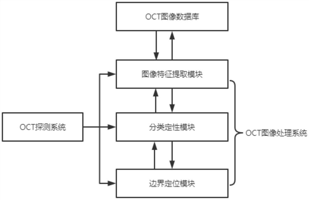
[0046] The image of the OCT image database is an image feature extraction module, the classification qualitative module, and the boundary positioning module processed by the OCT image processing system, and the depth fusion of multiple feature is performed by the classification qualitative module;

[0047] The comprehensiveness of the OCT image database also includes comparison the OCT image of different tissues to its histopathology;

[0048] The OCT image database is built, and includes the use of computer technology for feature extraction, classification qualitative, and boundary positioning of OCT image database;

[0049] Use Zeiss's 1300nm-band sweep frequency OCT system to collect two-dimensional / three-dimensional OCT images of animal living brain tissue and intraoperative human brain tissue, compare the OCT image of different tissues with its histopathology, as A portion of the OCT image database, while utilizing an OCT image pre-p...

Embodiment 2

[0051] OCT image characteristic extraction

[0052] The OCT image pre-processing module of the image feature extraction module is pre-processed from the enhanced contrast, image denoising and image registration;

[0053] The texture characteristics and depth features of the OCT image after the OCT image pre-processed are multi-feature extracted, the texture characteristics with the pre-processed OCT image as extraction object, extracts 5-dimensional straight diagram characteristics , 92 dimensional gray symbiotic matrix characteristics, 44 dimensional gray run length matrix characteristics; by extracting the ROI region grayscale matrix, calculating the grayscale histogram of the image, the extracted texture characteristic has a mean, variance, silakeity, peak and energy;

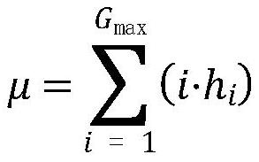
[0054] The mean calculation formula is as follows:

[0055]

[0056] Where H i For the pixel frequency of the grayscale value I, g max For the maximum gray value of the image;

[0057] Variance calculation for...

Embodiment 3

[0074] Subrigration of OCT images extracted with multiple feature

[0075] The classification qualitative module performs deep fusion of the above-mentioned multi-characterization, based on the SVM classifier core function mechanism, input depth features and texture features in multiple core functions and their multiple parameters, find the most suitable texture characteristics and depth features, respectively. The core function and parameter settings, and the weight of the respective nuclear functions, and then fuse the depth feature kernel function and the texture feature kernel function together to realize the multiple feature to perform multi-core function classification of multi-character species by the SVM classifier;

[0076] The image of multi-core function classification using the SVM classifier is characterized by comparing the OCT image database, and the OCT image database contrasts the multi-characterization of texture characteristics, depth features, and fusion;

[00...


PUM


Abstract

The invention discloses an OCT (Optical Coherence Tomography) image processing device for qualitative and boundary positioning in a brain tumor operation, which comprises an image feature extraction module and a feature extraction module, wherein the image feature extraction module comprises an OCT image preprocessing module and a feature extraction module for performing multiple feature extraction on textural features and depth features after OCT image preprocessing; the classification and qualitative module is used for performing deep fusion on the extracted multiple features, performing classification by adopting a feature fusion algorithm, and establishing a corresponding OCT image database; the method comprises the following steps: preprocessing an OCT image, performing multiple feature extraction on texture features and depth features of the OCT image after preprocessing the OCT image, obtaining a feature map of the image through pooling and convolution, expanding the feature map, connecting a full connection layer to realize final classification output, and performing automatic identification technology by mining deep information in the image to realize classification output of the OCT image. Doctors can be assisted to diagnose lesions and position boundaries; and performing feature extraction and classification of tissues and boundary positioning of brain glioma by combining an artificial image comparison and automatic identification technology.

Description

Technical field [0001] OCT images processing device and the qualitative brain tumor surgery The present invention relates border location for technical field involved in tumor imaging. Background technique [0002] Brain tumor is a growth of abnormal cells in the brain, according to the source of the abnormal cells generally can be divided into primary and secondary brain tumors, brain tumor types; primary brain tumors are derived directly from an abnormal growth of brain tissue cells, secondary brain tumors are diffused into the abnormal brain cells elsewhere in the body normal cells into cancer cells. Intraoperative qualitative and boundary locations of brain tumors is to assist the diagnosis and treatment of brain tumors in the image of the most important and most critical step, which can be a doctor's preoperative planning and intraoperative qualitative boundary positioning provide a reliable basis to ensure that brain tumor resection can more thoroughly, and do not harm norm...

Claims


Application Information

Patent Timeline
Patent Type & Authority Applications(China)
IPC IPC(8): G06T7/00G06V10/764G06V10/80G06V10/44G06V10/50G06V10/30G06K9/62G06N3/04G06N3/08
CPCG06T7/0012G06N3/08G06T2207/10101G06T2207/20081G06T2207/20084G06T2207/30096G06N3/045G06F18/2411G06F18/253
Inventor 杨建凯樊博戴丽刘红江杨松吕中强范振增
Owner THE SECOND HOSPITAL OF HEBEI MEDICAL UNIV
Why Eureka
Eureka Blog
Learn More