Chromosome image processing method and system

An image processing and chromosome technology, applied in the field of chromosome analysis, can solve the problems affecting the accuracy of automatic analysis of chromosome karyotypes, poor universality of multi-center images, and lack of uniform standards for image processing in chromosome metaphases, and achieves good generalization performance and universality. strong effect

Active Publication Date: 2022-03-25
易构智能科技(广州)有限公司
View PDF9 Cites 3 Cited by
  • Summary
  • Abstract
  • Description
  • Claims
  • Application Information

AI Technical Summary

Problems solved by technology

[0003] However, there is no uniform standard for the image processing of chromosome metaphase. The current commercial karyotype analysis systems or equipment (Leica, Zeiss, ASI equipment) all use self-developed special processing algorithms, resulting in the chromosome metaphase generated by different chromosome karyotype analysis systems. The image has a certain difference
In addition, in clinical work, different medical testing institutions also have differences in the links of training, production, and photography, and various factors lead to large differences in the chromosome metaphase images of different centers
The differences in these images lead to poor universality of automated algorithms for multi-center images, which in turn affects the accuracy of automated karyotype analysis

Method used


Image

  • Chromosome image processing method and system
  • Chromosome image processing method and system
  • Chromosome image processing method and system

Examples


Embodiment 1

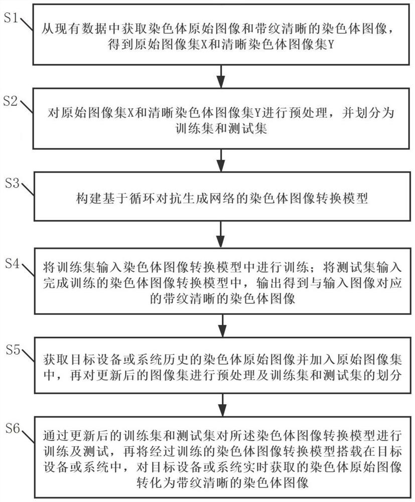
[0030] see Figure 1~2 , the present embodiment proposes a chromosome image processing method, which includes the following steps:

[0031] Step 1: Obtain the original chromosome image and the chromosome image with clear stripes from the existing data, and obtain the original image set X and the clear chromosome image set Y.

[0032] Step 2: Preprocess the original image set X and the clear chromosome image set Y, and divide them into a training set and a test set; the training set includes the original chromosome image and the chromosome image with clear stripes, and the test set includes the original chromosome image.

[0033] In a specific implementation process, the step of preprocessing the image includes: expanding the edge of the image to an image of a preset size, and then filling the content of the image after edge expansion to obtain an image of a uniform size, so as to realize the normalization of the image It can be processed without changing the shape and resolut...

Embodiment 2

[0050] In this embodiment, on the basis of the chromosome image processing method proposed in Embodiment 1, an image denoising processing step is added.

[0051] Specifically, the chromosome image processing method proposed in this embodiment further includes the following steps: performing denoising processing on the chromosome image with clear stripes output by the chromosome image conversion model, such as clump noise, cell debris noise, tissue fluid left to form Irregular flocculent or granular noise, as well as other cell chromosomes, chromosomes splashed over, and then a clean, clear chromosome image with stripes is obtained.

[0052] In a specific implementation process, a chromosome denoising model based on a semantic segmentation network is constructed to denoise the striated and clear chromosome image output by the chromosome image conversion model; wherein, the chromosome denoising model Each pixel in the input image is assigned a semantic category, the chromosome i...

Embodiment 3

[0058] see Figure 6 , this embodiment proposes a chromosome image processing system, which is applied to the chromosome image processing method proposed in Embodiment 1 or 2.

[0059] The chromosome image processing system proposed in this embodiment includes:

[0060] The collection module is used to collect the original chromosome image and the chromosome image with clear stripes;

[0061] The preprocessing module is used to normalize the collected original chromosome image and the chromosome image with clear stripes to obtain the preprocessed original image set X and the clear chromosome image set Y;

[0062] The chromosome image conversion module includes a chromosome image conversion model, and the chromosome image conversion model is trained through the original image set X and the clear chromosome image set Y;

[0063] The chromosome image conversion module is used to perform image conversion on the input original chromosome image, and output the corresponding chromo...


PUM


Abstract

The invention relates to the technical field of chromosome analysis, and provides a chromosome image processing method and system, and the method comprises the following steps: obtaining an original chromosome image and a clear chromosome image with stripes, and obtaining an original image set and a clear chromosome image set; preprocessing the image set, and dividing the image set into a training set and a test set; constructing a chromosome image conversion model based on a cyclic generative adversarial network; respectively inputting the training set and the test set into a chromosome image conversion model for training and testing; acquiring a historical chromosome original image of a target device or system, adding the historical chromosome original image into an original image set, preprocessing the updated image set, and dividing a training set and a test set; and training and testing the chromosome image conversion model through the updated training set and test set, carrying the trained chromosome image conversion model in a target device or system, and converting an original chromosome image obtained by the target device or system in real time into a chromosome image with clear stripes.

Description

technical field [0001] The present invention relates to the technical field of chromosome analysis, and more specifically, to a chromosome image processing method and system. Background technique [0002] Karyotype analysis is an important standard for the diagnosis of chromosomal abnormalities. The main process of chromosome karyotype analysis is to collect amniotic fluid, peripheral blood, and bone marrow, obtain metaphase samples through cell culture, staining, and film preparation, and then obtain metaphase images through digital photography, and finally analyze the metaphase images. When analyzing the karyotype of chromosome metaphase images, a clean and clearly striated image is the prerequisite for accurate analysis results. [0003] However, there is no uniform standard for the image processing of chromosome metaphase. The current commercial karyotype analysis systems or equipment (Leica, Zeiss, ASI equipment) all use self-developed special processing algorithms, re...

Claims


Application Information

Patent Timeline
Patent Type & Authority Applications(China)
IPC IPC(8): G06T7/00G06T5/00
CPCG06T7/0012G06T5/002G06T2207/20081G06T2207/30204
Inventor 李卫鹭
Owner 易构智能科技(广州)有限公司
Why Eureka
Try Eureka