Method and device for detecting artificial tissues and organoids

A technology of organoids and tissues, which is applied in the field of detection methods and devices of artificial tissues and organoids. It can solve the problems of small characteristic morphology, limited imaging penetration depth, difficult deep tissue imaging, etc., and achieve the effect of shortening the segmentation time.

Pending Publication Date: 2022-05-13
REGENOVO BIOTECH
View PDF0 Cites 2 Cited by
  • Summary
  • Abstract
  • Description
  • Claims
  • Application Information

AI Technical Summary

Problems solved by technology

However, due to the small characteristic morphological structure of artificial tissues and organoids, there is no obvious difference with the surrounding medium. With the prolongation of culture time, the change of the characteristic morphological structure cannot be observed with the naked eye. Therefore, it is necessary to segment and quantify the characteristic morphological structure Comparing the growth of artificial tissues and organoids to study the effect of a drug on a disease
[0003] Traditional detection methods are invasive, such as stained histology, fluorescence microscopy, bright field microscopy, etc., but cannot evaluate the real state of cells, and do not have volume structure imaging capabilities, and cannot quantify volume morphological characteristics, because such methods detect artificial Quantitative analysis of tissues or organoids is based on planar information or approximates artificial tissues or organoids as spheres or ellipsoids. However, the growth of organoids is irregular, which will cause large errors
At the same time, such methods have limited imaging penetration depth, making it difficult to perform deep tissue imaging.

Method used


Image

  • Method and device for detecting artificial tissues and organoids
  • Method and device for detecting artificial tissues and organoids
  • Method and device for detecting artificial tissues and organoids

Examples


Embodiment Construction

[0123] The present invention is further analyzed below in conjunction with specific examples.

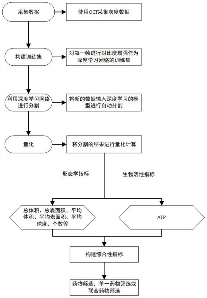
[0124] An OCT-based method for quantifying artificial tissues or organoids, see figure 1 , including the following steps:

[0125] Step (1): Using OCT equipment to collect the original three-dimensional grayscale image of the artificial tissue or organoid;

[0126] Open the OCT system, set the file format and storage path of the collected data, and set the file format according to the grayscale data to be collected. Select the acquisition mode in the software interface. The modes include 2D, 3D, Doppler and speckle. In this embodiment, the 3D mode is selected, and the field of view size, pixels and scanning speed of the acquisition are set in the 3D mode. The detected holes should be within the field of view, and the pixels should be set according to the required image clarity and data size. The default scanning speed is 48kHz. Ascan and Bscan do not need to be collected repeated...


PUM


Abstract

The invention discloses a method and a device for detecting artificial tissues and organoids. According to the method, artificial tissue and organ-like segmentation is realized by using an artificial tissue and organ-like segmentation deep learning network, and an artificial tissue and organ-like region three-dimensional image is obtained; the label adopts a contrast-enhanced image; calculating morphological indexes and ATP (adenosine triphosphate) values of artificial tissues and organoids in each culture hole of the corresponding culture plate in the last day of culture according to the connected domain; performing standardization processing on the morphological indexes of the artificial tissues and the organoids and the ATP numerical values to construct a matrix, and calculating a covariance matrix; performing singular value decomposition on the covariance matrix to obtain a right singular matrix and a singular value matrix; and main components are extracted according to the variance contribution rate, and artificial tissue and organoid comprehensive indexes are obtained. The comprehensive indexes of the artificial tissue and the organoid are used for researching the curative effect of the medicine.

Description

technical field [0001] The invention belongs to the field of detection of artificial tissues and organs, in particular to an OCT-based detection method and device for artificial tissues and organoids. Background technique [0002] Artificial tissues and organoids are 3D cell complexes formed by self-assembly driven by human-derived cells or stem cells, which can simulate the structure and function of human organs, and can be used for optimal screening of drugs, pathological exploration, etc., in artificial tissues and In the process of organoid culture or drug screening of artificial tissues and organoids with specific physiological structures, the morphological indicators of artificial tissues or organoids are closely related to their functional status. Through high-resolution continuous morphological observation, artificial tissues or organoids can be detected. Growth status of organoids. However, due to the small characteristic morphological structure of artificial tissu...

Claims


Application Information

Patent Timeline
Patent Type & Authority Applications(China)
IPC IPC(8): G06T7/00G06T7/11G06T5/00G06T5/30G06N3/04G06N3/08
CPCG06T7/0012G06T7/11G06T5/002G06T5/007G06T5/30G06N3/08G06T2207/10101G06T2207/20036G06T2207/20081G06T2207/20084G06T2207/30028G06T2207/30096G06N3/045
Inventor 王玲张琳翊杨珊珊徐铭恩
Owner REGENOVO BIOTECH
Why Eureka
Try Eureka