Eureka AIR delivers breakthrough ideas for toughest innovation challenges, trusted by R&D personnel around the world.

Imaging method and device applied to cone beam CT sparse sampling

A technology of sparse sampling and imaging method, applied in the field of medical imaging, can solve the problem of slow image processing speed, and achieve the effect of amplifying fidelity, enhancing fidelity, and improving the signal-to-noise ratio of reconstructed images

Pending Publication Date: 2022-05-17
BEIJING CANCER HOSPITAL PEKING UNIV CANCER HOSPITAL
View PDF0 Cites 2 Cited by
  • Summary
  • Abstract
  • Description
  • Claims
  • Application Information

AI Technical Summary

Problems solved by technology

However, the image processing speed of this technical solution is still slow, and it is difficult to perform faster processing on the imaging of different body parts of the patient.

Method used


Image

  • Imaging method and device applied to cone beam CT sparse sampling
  • Imaging method and device applied to cone beam CT sparse sampling
  • Imaging method and device applied to cone beam CT sparse sampling

Examples


Embodiment Construction

[0032] In order to make the object, technical solution and advantages of the present invention clearer, the present invention will be described in further detail below in conjunction with specific embodiments and with reference to the accompanying drawings.

[0033]It should be noted that, in the accompanying drawings or in the text of the specification, implementations that are not shown or described are forms known to those of ordinary skill in the art, and are not described in detail. In addition, the above definitions of each element and method are not limited to the various specific structures, shapes or methods mentioned in the embodiments, and those skilled in the art can easily modify or replace them.

[0034] It should also be noted that the directional terms mentioned in the embodiments, such as "up", "down", "front", "back", "left", "right", etc., are only referring to the directions of the drawings, not Used to limit the protection scope of this disclosure. Throug...


PUM


Abstract

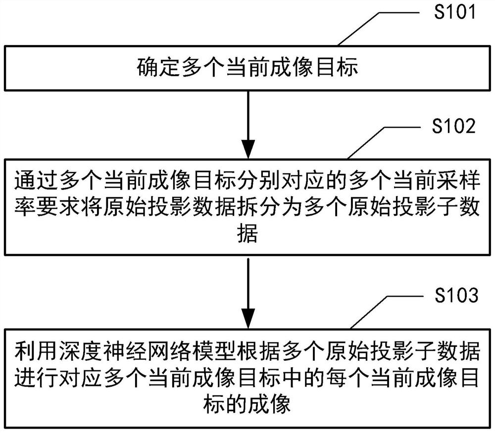
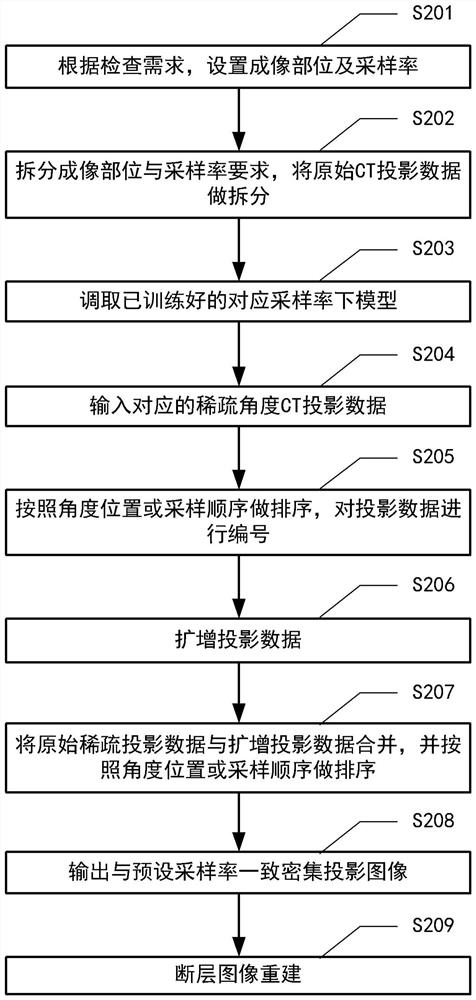
The invention provides an imaging method and device applied to cone beam CT sparse sampling, a medium and a program product. The imaging method applied to cone beam CT sparse sampling comprises the following steps: determining a plurality of current imaging targets; splitting the original projection data into a plurality of original projection sub-data according to a plurality of current sampling rate requirements respectively corresponding to the plurality of current imaging targets; and performing imaging corresponding to each current imaging target in the plurality of current imaging targets according to the plurality of original projection sub-data by using a deep neural network model. Compared with an existing traditional scheme which can cause the problem of error amplification through a cyclic interpolation mode, the method can directly amplify subdivision position projection, meanwhile, a residual error and structural similarity double-constraint neural network model is combined, the fidelity of an amplified projection image is enhanced, the imaging speed and the image processing speed are greatly improved, and the image processing efficiency is improved. And under the condition of meeting low-dose irradiation, the signal-to-noise ratio, the structural similarity and the overall image quality of the reconstructed image are remarkably improved.

Description

technical field [0001] The present disclosure relates to the technical field of medical imaging, and in particular to an imaging method, device, medium and program product applied to cone-beam CT sparse sampling. Background technique [0002] Computed tomography (CT) is one of the key imaging technologies in the field of medical imaging, widely used in radiology, oral imaging, image-guided surgery, image-guided radiotherapy and other fields. CT imaging requires X-rays, and excessive X-ray radiation will bring safety risks to human health. Therefore, low-dose CT has great health significance and is also one of the important directions for the development of CT technology. [0003] Cone-Beam Compute Tomography (CBCT for short) onboard a radiotherapy accelerator, as a key supporting technology for Image-guided Radio Therapy (IGRT for short), can not only quickly provide three-dimensional tomographic images of patients, but also can Six-dimensional (6Degree-of-Freedom, referred...

Claims


Application Information

Patent Timeline
Patent Type & Authority Applications(China)
IPC IPC(8): G06T7/00G06N3/04G06N3/08
CPCG06T7/0012G06N3/08G06T2207/10081G06T2207/20081G06N3/045
Inventor 杜乙王若曦赵旭之岳海振李丹妮朱向高吴昊彭亚辉
Owner BEIJING CANCER HOSPITAL PEKING UNIV CANCER HOSPITAL
Why Eureka
Eureka Blog
Learn More