
Split iliac vein stent and placement method thereof

A technology for iliac veins and veins, applied in the field of split iliac vein stents and their placement, which can solve the problems of difficulty in determining the placement position of the stent, difficult operation, and complicated release process of the stent, and achieve easy determination of the placement position, reduced operation difficulty, Avoid the effects of blood flow disturbance

Pending Publication Date: 2022-05-27
WEST CHINA HOSPITAL SICHUAN UNIV
View PDF6 Cites 0 Cited by
  • Summary
  • Abstract
  • Description
  • Claims
  • Application Information

AI Technical Summary

Problems solved by technology

The former can be accurately positioned and placed in the common iliac vein, but lacks flexibility, cannot cross the hip joint, and cannot adhere well to the wall
The latter is prone to shortening and poor support
The main problems faced by the existing stents are: (1) during the surgical implantation of the stent, the placement position of the stent is difficult to determine, the release process of the stent is more complicated, and the operation is difficult; (2) to ensure complete coverage of common iliac vein lesions, the stent It needs to be partially released into the inferior vena cava, which will change the original blood flow direction, shape of the blood flow cavity and blood flow field. Lead to the formation of thrombus; ③ Postoperative stents may have displacement problems
However, the placement method of the Y-shaped stent is much more complicated than the existing stents, and the clinical operation is difficult, and the main stent of the inferior vena cava section will increase the burden on the vein wall, which is easy to cause damage to the vein wall, and may even cause in-stent failure. Waiting for new questions

Method used


Image

  • Split iliac vein stent and placement method thereof
  • Split iliac vein stent and placement method thereof
  • Split iliac vein stent and placement method thereof

Examples


Embodiment 1

[0035] The diseased veins are mainly due to iliac vein compression syndrome (Iliacvenous compression syndrome, IVCS for short). IVCS is a disease of lower extremity and pelvic venous return obstruction caused by iliac vein compression and / or the existence of abnormal adhesion structures in the cavity. Usually, the patient's right common iliac artery crosses the left common iliac vein, which is easy to cause external compression of the vein, resulting in iliac vein occlusion or stenosis. This compression and intraluminal changes predispose the patient to left lower extremity deep vein thrombosis. Failure to recognize and treat this abnormality in patients with acute thrombosis can lead to severe vascular sequelae and chronic left leg symptoms. This kind of thromboembolism is difficult to recanalize, and can cause venous return disorders in the lower extremities and pelvis and lower extremity venous hypertension.

[0036] In order to solve the problems in endovascular intervent...

Embodiment 2

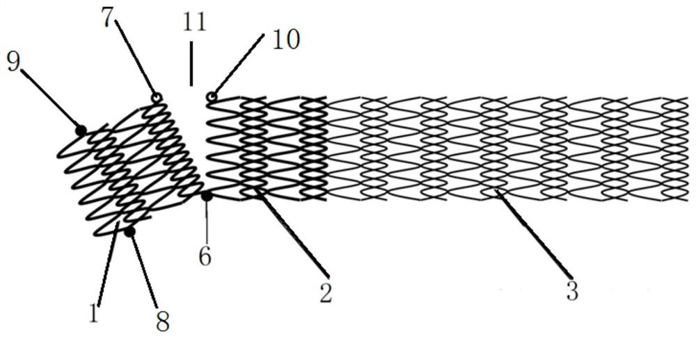
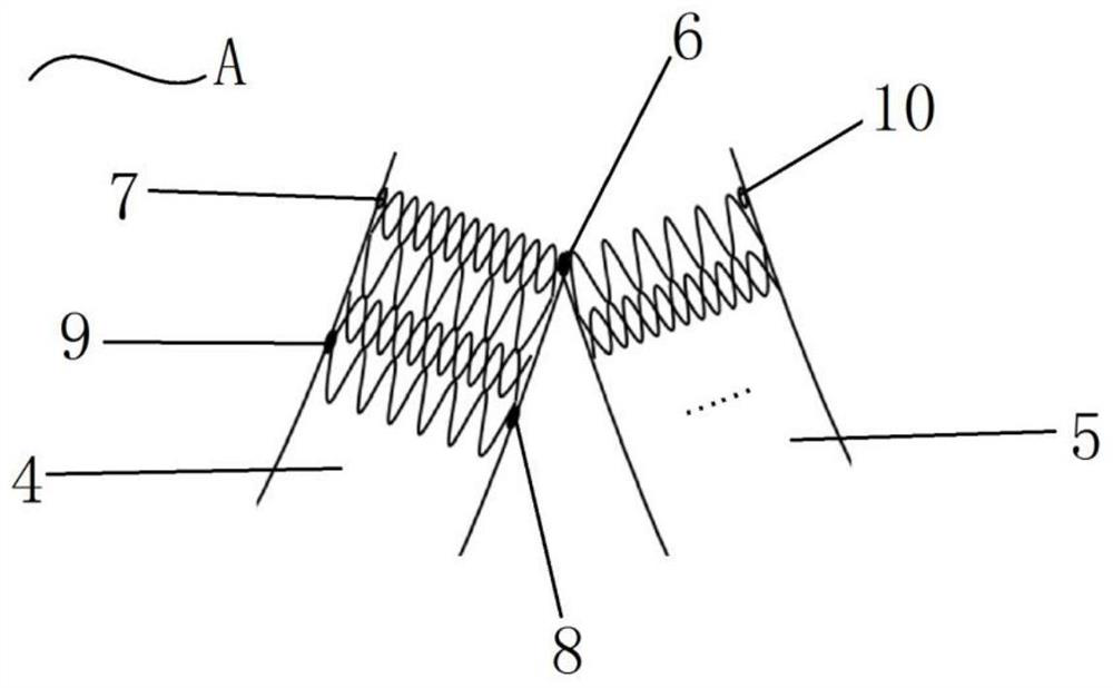
[0040] The difference between Embodiment 2 and Embodiment 1 is that a split iliac vein stent includes a distal segment 2 and a proximal segment 3 of the diseased vein that are connected to each other in the diseased vein, and a stent located in another common iliac vein. The segment 1 opposite to the diseased vein, the segment 1 opposite to the diseased vein and the distal segment 2 of the diseased vein can be divided by a stent, and the dividing part is not completely disconnected to form a split opening 11, and the connecting part is formed at the place that is not disconnected, At this time, the connecting part is any arc on the second end of the distal segment of the diseased vein or the segment 1 on the opposite side of the diseased vein, and the arc length of the connecting part is 4-7 mm (or about the distal segment 2 of the diseased vein). 1 / 8-1 / 6 of the perimeter of the end section), and a first developing point 6 is provided at the midpoint of the arc length of the co...

Embodiment 3

[0044] The difference between Embodiment 3 and Embodiment 1 is that a split iliac vein stent in this embodiment includes a distal segment 2 of the diseased vein and a proximal segment 3 of the diseased vein that are connected to each other in the diseased vein, and another iliac vein located in the other iliac vein. The diseased vein contralateral segment 1 of the common vein, the diseased vein is one of the two common iliac veins. In this embodiment, the diameters of the segment 1 on the opposite side of the diseased vein and the segment 2 at the distal end of the diseased vein may be different, and the adjustment method depends on the specific conditions of the patient; for example, the diameter of the segment 1 on the opposite side of the diseased vein is 12 mm and the length is 25 mm , the diameter of the distal segment 2 of the diseased vein is 15mm and the length is 20mm; the diameter of the proximal segment 3 of the diseased vein is 15mm and the length is 30mm. The conn...


PUM

PropertyMeasurementUnit
Arc lengthaaaaaaaaaa
Diameteraaaaaaaaaa
Lengthaaaaaaaaaa

Abstract

The invention discloses a split-type iliac vein stent and a placement method thereof.The split-type iliac vein stent comprises a lesion vein far-end section and a lesion vein near-end section which are located in a lesion vein and connected with each other and a lesion vein opposite-side section located in another iliac total vein, a connecting part is arranged at one end of the lesion vein far-end section, a first developing point is arranged on the connecting part, and a second developing point is arranged at the other end of the lesion vein near-end section; the lesion vein far-end section is connected with one end of the lesion vein opposite-side section through the connecting part, the lesion vein opposite-side section and the lesion vein far-end section form an inverted V shape at the convergence position of the two iliac total veins, and the lesion vein and the opposite side of the lesion vein are connected in a separated mode, so that the lesion vein and the opposite side of the lesion vein can be connected in the blood flowing direction. The stent is split at the intersection of the left and right iliac general veins to form a V-shaped structure, so that the stent is matched with the special form of the convergence position of the left and right iliac general veins to avoid displacement, the stent cannot enter the inferior vena cava, and the mode is convenient for medical staff to position and place and easy to operate.

Description

technical field [0001] The invention relates to the technical field of medical devices, in particular to a split iliac vein stent and a placement method thereof. Background technique [0002] At present, in order to restore the patency of the iliac veins, the treatment of iliac vein compression syndrome mainly adopts endovascular interventional surgery, which is mainly to perform stent implantation or balloon dilation at the location of iliac vein lesions, which is simple, minimally invasive, and remote. The patency rate is high during the stent implantation operation; the stent is usually a hollow or braided tubular structure, which can be used for the recanalization of pathologically changed hollow organs. The stent in the compressed state can be introduced into the position to be treated through the delivery catheter, and certain measures can be taken to expand it, so as to achieve the supporting effect. Although there are many types of vascular stents on the market, the...

Claims


Application Information

Patent Timeline
IPC IPC(8): A61F2/86
CPCA61F2/86
Inventor 袁丁曹皓瑶郑庭辉王家嵘
Owner WEST CHINA HOSPITAL SICHUAN UNIV
Learn More