

Method of predicting endometrial receptivity
A technology of endometrium and receptivity, which is applied in the field of prediction of endometrial receptivity, and can solve problems such as inability to accurately determine specific participation
- Summary
- Abstract
- Description
- Claims
- Application Information
AI Technical Summary
Problems solved by technology
Method used
Image

Examples
Embodiment 1
[0344] Example 1: Materials and Methods
[0345] Human endometrial tissue for isolation of primary endometrial epithelial cells
[0346] Ethical approval was obtained from the Human Ethics Committee at Monash Medical Centre (Melbourne, Australia) and all patients provided informed written consent. Endometrial biopsy samples were obtained from women who had undergone hysteroscopic dilation, curettage, or assessment of tubal patency. Menstrual cycle stages were determined by routine histological dates of tissues.
[0347] Isolation of Primary Human Endometrial Epithelial Cells (HEEC)
[0348] Tissues in the proliferative phase (days 6-14) were harvested into Dulbecco's modified Eagle's medium / F12 (DMEM / F12, Thermo Fisher Scientific, MA, USA) and cells were detached within 24 hours of collection. Cells were isolated by enzymatic digestion and filtration as previously described (Marwood et al., 2009). Briefly, collagenase (7.5 U / ml; Sigma) and DNase 1 (2000 U / ml; Roche, Castle...
Embodiment 2
[0411] Example 2: Proteomic identification of podocalyx proteins in primary human endometrial epithelial cells
[0412] As described in Example 1, primary endometrial epithelial cells (HEECs) from human endometrial tissue were isolated and enriched for plasma membrane proteins.
[0413] A total of 250 proteins were identified by mass spectrometry analysis of the obtained proteins (Table 2). Of these, 47 are considered to be cell membrane proteins, and 10 of them are involved in cell adhesion, including podocalyx protein (PCX).
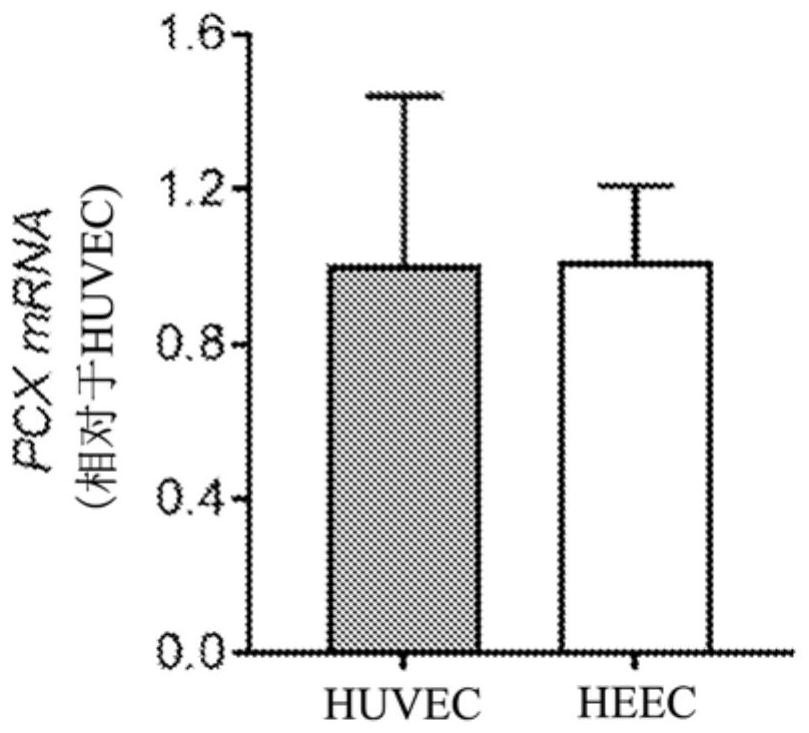
[0414] To confirm the proteomic findings, total cell lysates of primary HEEC isolated from proliferating endometrium (for proteomic studies) were analyzed by Western blot using 3 antibodies directed against different regions of human PCX.
[0415] A dominant band of about 150 kDa was detected by all 3 antibodies, with a level of compatibility in both cell types. Ab1 detected an additional weaker band of about 80 kDa in HUVEC and HEEC, while Ab2 recog...
Embodiment 3
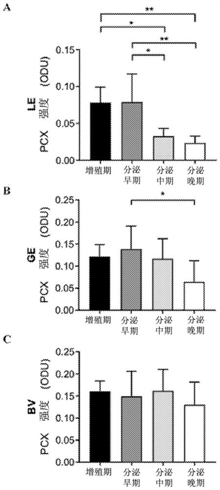
[0435] Example 3: PCX localizes to the apical membrane of human endometrial epithelial cells and endothelial cells and is specifically downregulated in the luminal epithelium, consistent with receptivity establishment
[0436] As described in Example 1, the cellular localization of PCX in the human endometrium throughout the menstrual cycle was examined by immunohistochemistry.
[0437] Similar staining patterns were detected for all 3 PCX antibodies. During the proliferative phase, PCX is strongly localized to the apical surface of luminal and glandular epithelial cells (LE and GE, respectively) and endothelial cells in blood vessels (BV). The matrix shows no / under detection. This pattern persisted more or less until early secretion, after which large differences emerged, especially in LE. In mid-secretory phase, PCX staining was barely detectable in LE, although still strong in both GE and BV. During late secretion, while LE continued to have minimal PCX, GE showed lighte...
PUM
 Abstract
Description
Claims
Application Information