Homologous recombination defect detection method

A defect detection and homologous recombination technology, applied in biochemical equipment and methods, microbial determination/inspection, etc., can solve problems such as insufficiency, inability, and large amount of data

Pending Publication Date: 2022-07-01
YIKON GENOMICS SHANGHAI CO LTD
View PDF0 Cites 0 Cited by
  • Summary
  • Abstract
  • Description
  • Claims
  • Application Information

AI Technical Summary

Problems solved by technology

However, the current domestic HRD status assessment products all use the next-generation sequencing method. 1. This method requires a relatively high initial sample volume (above 250ng); 2. This method generally selects approximately There are 70,000 SNPs, the captured interval is about 2M, and the amount of sequencing data is large. The cost of detection reagents for a sample is more than 1,000 for the capture of SNP sites and sequencing, plus the operating cost of the detection methodology. The clinical fee is about 10,000 yuan, and the high cost problem is the pain point of widespread clinical application, which greatly reduces the number of people who benefit from this test, and cannot become an effective screening method for companion diagnosis; 3. Can not meet the requirements at the same time The detection of other drugs can not meet the demands of doctors and patients clinically; 4. The detection cycle of NGS generally takes 6-7 days, which also makes patients need a long waiting time

Method used


Image

  • Homologous recombination defect detection method
  • Homologous recombination defect detection method
  • Homologous recombination defect detection method

Examples


Embodiment 1

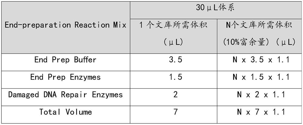
[0040] Example 1: Total DNA extraction

[0041] For a puncture sample, use the QIAamp DNA FFPE Tissue Kit (QIAGEN, Cat. No. 56404) to extract the FFPE paraffin sample. After extraction, 25ng-200ng will be obtained, of which 200ng will be taken out for DNA fragmentation.

Embodiment 2

[0042] Example 2: DNA Fragmentation

[0043] 1. Set up Covaris S220

[0044] Pour fresh deionized water into the sink until level = 12 (Covaris S220). Turn on the power switches of the Covaris, the cooling unit and the circulation system in sequence. If the loop system front panel shows OFF, press and hold the Enter key to turn the switch on. Set the cooling device to 4°C. Deionized water needs to be vented for at least 30 minutes. Keep the pump on at all times during sample processing.

[0045] 2. Prepare the samples that need to be interrupted

[0046] In a 1.5mL low adsorption tube, dilute different concentrations of sample gDNA or FFPE DNA to a volume of 52.5μL with 1x Low TE, taking the maximum value in the table below, vortex to mix, and centrifuge briefly.

[0047] DNA concentration DNA Quality Rating Total Input (ng) DNA concentration ≥ 1.2 A 65-220 DNA concentration ≥ 1.2 B 65-330 DNA concentration ≥ 2.5 C 130-440

[0048] ...

Embodiment 3

[0065] Example 3: Joint Connection

[0066] 1. Connector connection

[0067] Take out the reagents Ligation Buffer, Ligation Enzymes and UDIadapters from the kit in the -20°C refrigerator. The Buffer and adapter are thawed at room temperature, vortexed and mixed, then centrifuged briefly and placed on ice. After the enzyme is flicked, centrifuged briefly and placed on ice. Note that different indexes are selected for libraries built in the same batch.

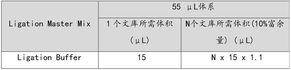
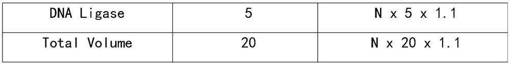
[0068] Prepare Ligation's Master Mix in a 1.5 mL sterile tube and operate on ice.

[0069]

[0070]

[0071] Set the program on the PCR machine in advance, 60min at 20℃, hold at 4℃, named Ligation.

[0072] NOTE: The heated lid temperature is set to 103°C.

[0073] 20℃ 60min 4℃ Hold

[0074] To each end-repaired 0.2 mL PCR tube, add 20 μL of Ligation Master mix. Add 5 μL of the corresponding Adapter to each tube, mix by slowly pipetting 10 times with a pipette, and centrifuge briefly until the fi...


PUM


Abstract

According to the homologous recombination defect detection method provided by the invention, the detection of accompanying diagnosis of multiple drugs can be simultaneously met, the requirement on the initial quantity of a sample is low, the cost is low, and the detection period is short; the homologous recombination defect detection method comprises the following steps: S1, carrying out total DNA extraction, DNA fragmentation and DNA amplification on a human tissue sample; s2, detecting a part of amplified tissue sample DNA by adopting an SNP chip, and calculating an HRD value; next-generation sequencing is carried out on the other part of amplified tissue sample DNA, and the gene mutation state in an HRD signal channel and accompanying diagnosis of other targeted drugs are evaluated; and S3, integrating an SNP chip detection result and a next-generation sequencing result, and judging which tumor treatment medicine the sample can benefit from.

Description

technical field [0001] The present invention relates to the field of biotechnology, and more particularly, to a method for detecting homologous recombination deficiency. Background technique [0002] For the detection of HRD (homologous recombination deficiency, homologous recombination deficiency), the total amount of tissue samples from tumor patients is very limited. Depending on the diameter of the puncture needle, the tumor tissue obtained by one-time puncture ranges from tens of thousands of cells to millions of cells. With the development of precision medicine, the detection methodologies that need to be done downstream have also expanded from one to many: for example NGS, RNAseq, microarrays, next-generation sequencing, ddPCR, nanostring, and more. However, the total number of clinical samples is too small, so clinicians often have to make difficult choices between different methodologies (corresponding to different medication guidance), so that patients are forced ...

Claims


Application Information

Patent Timeline
Patent Type & Authority Applications(China)
IPC IPC(8): C12Q1/6837C12Q1/6827C12Q1/6858C12Q1/6886
CPCC12Q1/6837C12Q1/6827C12Q1/6858C12Q1/6886C12Q2600/136C12Q2600/156C12Q2531/113C12Q2565/501C12Q2535/122C12Q2537/165
Inventor 程华石伟杰
Owner YIKON GENOMICS SHANGHAI CO LTD
Why Eureka
Try Eureka