Prostate image segmentation method and prostate cancer intelligent auxiliary diagnosis system

An image segmentation, prostate technology, applied in image analysis, image data processing, instruments, etc., can solve the problems of only detecting tumors close to the posterior capsule of the prostate, time-consuming labor, prostate cancer errors, etc., to achieve optimal classification performance, enabling identification and marking, and improving accuracy

Pending Publication Date: 2022-07-29
GUANGDONG UNIV OF TECH +1
View PDF0 Cites 4 Cited by
  • Summary
  • Abstract
  • Description
  • Claims
  • Application Information

AI Technical Summary

Problems solved by technology

However, the test level of serum PSA is affected by many factors in actual detection, such as digital rectal examination, indwelling catheter, prostatitis, sexual life and use of finasteride in patients with prostate cancer. Therefore, there are considerable errors and clinical missed diagnoses in predicting the detection rate of prostate cancer when the detection of serum prostate cancer-specific antigen is used alone in clinical practice.
Although the operation of digital rectal examination is simple and noninvasive, it is mainly judged subjectively, and only tumors close to the posterior capsule of the prostate can be detected during the exploration process. Therefore, in the diagnosis of prostate cancer In diagnosis, digital rectal examination also has considerable limitations in the clinical detection of prostate cancer
[0004] At the same time, since mpMRI of prostate cancer has a large amount of image data, the diagnosis process requires manual identification by doctors, which consumes a lot of time and labor. In addition, manual diagnosis results are subjective and are often limited by the experience of doctors, resulting in misdiagnosis

Method used


Image

  • Prostate image segmentation method and prostate cancer intelligent auxiliary diagnosis system
  • Prostate image segmentation method and prostate cancer intelligent auxiliary diagnosis system
  • Prostate image segmentation method and prostate cancer intelligent auxiliary diagnosis system

Examples


Embodiment 1

[0063] The purpose of this embodiment is to provide a prostate image segmentation method.

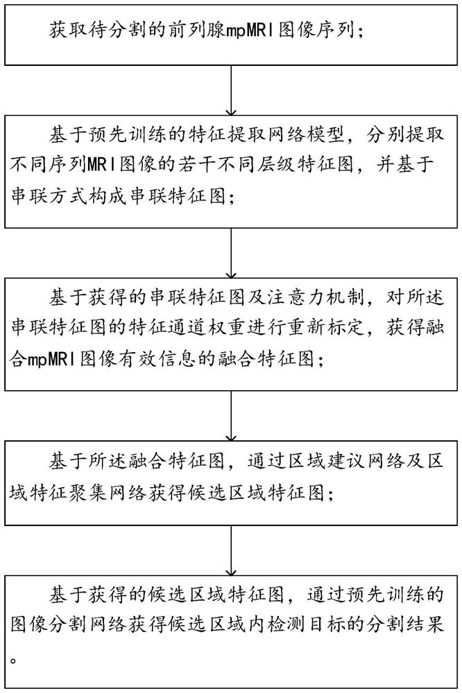
[0064] like figure 1 As shown, a prostate image segmentation method is provided, comprising:

[0065] Obtain the prostate mpMRI image sequence to be segmented;

[0066] Based on the pre-trained feature extraction network model, several feature maps of different levels of different sequences of MRI images are extracted respectively, and the serial feature maps are formed based on the concatenation method;

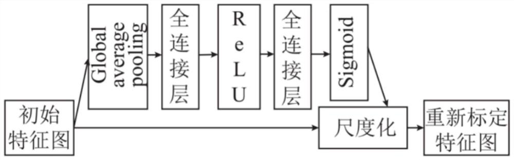
[0067] Based on the obtained tandem feature map and the attention mechanism, re-calibrate the feature channel weights of the tandem feature map to obtain a fusion feature map that fuses the effective information of the mpMRI image;

[0068] Based on the fusion feature map, obtain the candidate region feature map through the region proposal network and the region feature aggregation network;

[0069] Based on the obtained feature maps of the candidate regions, the segmentation resul...

Embodiment 2

[0093] The purpose of this embodiment is to provide an intelligent auxiliary diagnosis system for prostate cancer.

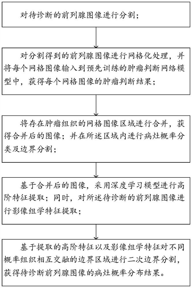
[0094] like figure 2 As shown, an intelligent auxiliary diagnosis system for prostate cancer is provided, including:

[0095] Segmenting the prostate image to be diagnosed, wherein the segmentation method adopts the above-mentioned prostate image segmentation method;

[0096] Perform grid processing on the segmented prostate images, and input each grid image into the pre-trained tumor judgment network model to obtain the tumor judgment result of each grid image;

[0097] Merging the grid image areas with tumor tissue to obtain a merged image; and performing lesion probability classification and boundary segmentation in the area;

[0098] Based on the merged images, a deep learning model is used to perform high-level feature extraction; at the same time, radiomics feature extraction is performed on the prostate image to be diagnosed;

[0099] Based on the ext...

Embodiment 3

[0125] The purpose of this embodiment is to provide an electronic device.

[0126] An electronic device, comprising a memory, a processor and a computer program stored on the memory to run, the processor implements the following steps when executing the program:

[0127] Segmenting the prostate image to be diagnosed, wherein the segmentation method adopts the above-mentioned prostate image segmentation method;

[0128] Perform grid processing on the segmented prostate images, and input each grid image into the pre-trained tumor judgment network model to obtain the tumor judgment result of each grid image;

[0129] Merging the grid image areas with tumor tissue to obtain a merged image; and performing lesion probability classification and boundary segmentation in the area;

[0130] Based on the merged images, a deep learning model is used to perform high-level feature extraction; at the same time, radiomics feature extraction is performed on the prostate image to be diagnosed;...


PUM


Abstract

The invention provides a prostate image segmentation method and a prostate cancer intelligent auxiliary diagnosis system, and belongs to the technical field of prostate cancer intelligent auxiliary diagnosis, the prostate image segmentation method comprises the following steps: segmenting a prostate image to be diagnosed by using the prostate image segmentation method; performing meshing processing on the prostate images obtained by segmentation, and inputting the processed prostate images into a pre-trained tumor judgment network model to obtain a tumor judgment result of each grid image; combining the grid images with the tumor tissues to obtain a combined image; lesion probability classification and boundary segmentation are carried out in the region; based on the merged image, performing high-order feature extraction by using a deep learning model; meanwhile, image omics feature extraction is carried out on the prostate image to be diagnosed; and based on the extracted high-order features and radiomics features, carrying out secondary boundary segmentation on a boundary region where tissues with different probabilities are mutually fused to obtain a lesion probability distribution result of the prostate image.

Description

technical field [0001] The disclosure belongs to the technical field of intelligent auxiliary diagnosis of prostate cancer, and in particular relates to a prostate image segmentation method and an intelligent auxiliary diagnosis system for prostate cancer. Background technique [0002] The statements in this section merely provide background information related to the present disclosure and do not necessarily constitute prior art. [0003] At present, the main clinical screening methods for prostate cancer are serum prostate-specific antigen detection and digital rectal examination. However, the test level of serum PSA is affected by various factors in actual detection, such as digital rectal examination, indwelling catheter, prostatitis, sexual life, and use of finasteride in patients with prostate cancer. Therefore, the detection of serum prostate cancer-specific antigen alone in clinical practice has considerable errors and clinical missed diagnosis in predicting the det...

Claims


Application Information

Patent Timeline
Patent Type & Authority Applications(China)
IPC IPC(8): G06T7/00G06V10/44G06V10/26G06V10/80G06K9/62G06N3/04
CPCG06T7/0012G06T2207/10016G06T2207/10088G06T2207/20081G06T2207/20084G06T2207/30081G06T2207/30096G06N3/048G06N3/045G06F18/253
Inventor 任鸿儒吴斌魏强吕世栋鲁仁全
Owner GUANGDONG UNIV OF TECH
Why Eureka
Try Eureka