
Left atrial appendage occlusion simulation system based on fusion of echocardiography and CT (Computed Tomography) multi-modal image

An echocardiography and multimodal image technology, applied in the field of medical devices, can solve the problems of failure, hasty, and falling off of left atrial appendage occlusion, and achieve the effect of improving the success rate of surgery, reducing complications, and improving the success rate.

Pending Publication Date: 2022-08-02
XIN HUA HOSPITAL AFFILIATED TO SHANGHAI JIAO TONG UNIV SCHOOL OF MEDICINE
View PDF0 Cites 0 Cited by
  • Summary
  • Abstract
  • Description
  • Claims
  • Application Information

AI Technical Summary

Problems solved by technology

[0003] However, with the widespread development of left atrial appendage occlusion, especially minimalist surgical methods, on the one hand, the high residual shunt rate after left atrial appendage occlusion is still one of the important problems faced by cardiologists. The risk of thrombus formation on the surface of the occluder increases in patients with a large number of patients. In addition to long-term anticoagulant therapy, the residual cavity needs to be re-occluded; The evaluation of the LAA and its adjacent shape is limited. During LAA occlusion, the diameter of the anchoring area and the depth of the LAA can also be measured, but the evaluation of the LAA is limited during the operation. hasty, imprecise
The above situation may lead to the failure of left atrial appendage occlusion, or the occurrence of more residual shunt flow after occlusion, occluder displacement or even falling off, etc. Similarly, surgeons also have difficulties in choosing occluder devices, which will affect the surgical effect

Method used


Image

  • Left atrial appendage occlusion simulation system based on fusion of echocardiography and CT (Computed Tomography) multi-modal image
  • Left atrial appendage occlusion simulation system based on fusion of echocardiography and CT (Computed Tomography) multi-modal image
  • Left atrial appendage occlusion simulation system based on fusion of echocardiography and CT (Computed Tomography) multi-modal image

Examples


Embodiment Construction

[0031] In order to make the present invention more obvious and easy to understand, preferred embodiments are hereby described in detail with the accompanying drawings as follows:

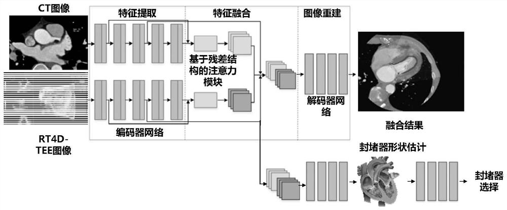
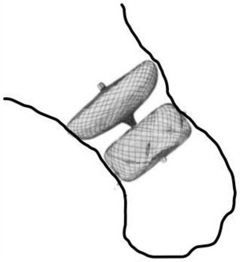
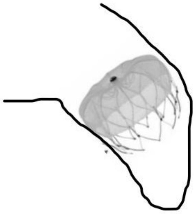
[0032] like Figure 1-3 As shown, in order to reduce the residual shunt rate after left atrial appendage occlusion and improve the success rate of surgery, the present invention provides a method for predicting the effect of left atrial appendage occlusion by fusion of real-time four-dimensional echocardiography and CT multimodal images through the esophagus, including the following step:

[0033] Step 1: Obtain the DICOM format image data of human left atrial appendage transesophageal real-time four-dimensional echocardiography and CT;

[0034] Step 2: Perform threshold segmentation on the CT images of the human heart; for typical cardiac segmentation tasks, the input CT images are generated by the full convolutional neural network (FCN) to generate right ventricle / left ventricle / right atrium / left...


PUM


Abstract

The invention relates to an echocardiography and CT multi-modal image fused left auricle occlusion simulation system, and belongs to the technical field of medical apparatuses and instruments. Through combined application of two imaging methods, DICOM image data of the two methods are collected respectively, software is designed to achieve fusion imaging, the internal structure, the function and the external form of the left atrial appendage are displayed at the same time, matching of a plugging device is simulated, and the left atrial appendage is simulated to be close to the left atrial appendage. According to the left auricle occlusion simulation device, a surgeon can select an operation strategy by using the left auricle occlusion simulation device before an operation, the optimal and most suitable occlusion device is selected, and the success rate of the operation is improved. The image fusion simulation system provided by the invention can realize fusion of a transesophageal real-time four-dimensional echocardiogram and a CT (Computed Tomography) multi-modal image of the left auricle, and performs fusion of an internal structure and functions and an external overall form of the left auricle and adjoining of the left auricle, so as to improve the success rate of left auricle occlusion. And a feasible imaging method is provided for reducing complications.

Description

technical field [0001] The invention relates to a left atrial appendage occlusion simulation system integrating echocardiography and CT multimodal images, and belongs to the technical field of medical devices. Background technique [0002] At present, in LAA occlusion surgery, the clinical application is mostly based on the guidance of transesophageal echocardiography or CT single modality imaging method, combined with intraoperative cardiovascular angiography to observe the effect of LAA occlusion. [0003] However, with the widespread development of left atrial appendage occlusion, especially minimalist surgical methods, on the one hand, the residual shunt rate after left atrial appendage occlusion is high, which is still one of the important problems faced by cardiologists. Many patients have an increased risk of thrombosis on the surface of the occluder, and in addition to long-term anticoagulation therapy, they also need to re-occlude the residual cavity; The evaluatio...

Claims


Application Information

Patent Timeline
Patent Type & Authority Applications(China)
IPC IPC(8): A61B34/10
CPCA61B34/10A61B2034/104
Inventor 郁怡顾运李毅刚杨杰汤晓黎潘小勇王倩黄晓红王婷陈牧
Owner XIN HUA HOSPITAL AFFILIATED TO SHANGHAI JIAO TONG UNIV SCHOOL OF MEDICINE
Learn More