
Layered osteotomy method by reference of superpose images

An osteotomy and image technology, which is applied to the image superimposed layered osteotomy method involving the medical field, can solve the problems of easy backward recovery, easy secondary infection, dead space in large bones, etc., so as to reduce blindness and reduce medical treatment. The effect of disputes

Inactive Publication Date: 2004-10-27
上海第二医科大学附属第九人民医院
View PDF0 Cites 12 Cited by
  • Summary
  • Abstract
  • Description
  • Claims
  • Application Information

AI Technical Summary

Problems solved by technology

The existing problem is that the entire orbitozygomatic outer skeleton is pulled by the craniofacial muscles after osteotomy and moves forward, so it is easy to recover backwards, and the shape effect is difficult to maintain for a long time; cavities, susceptible to secondary infection
[0006] In China, there are few reports on the application of computer-aided image texture segmentation based on craniofacial bone structure CT data, image extraction and reconstruction based on image analysis features, and calculation of volume changes after changes in the position of related bone structures in complex craniofacial deformities.
There is no report on the application of craniofacial layered osteotomy in complex craniofacial deformities

Method used


Image

  • Layered osteotomy method by reference of superpose images
  • Layered osteotomy method by reference of superpose images
  • Layered osteotomy method by reference of superpose images

Examples


Embodiment Construction

[0015] Design of three-dimensional overlay of orbital volume and its application in orbital osteotomy and displacement

[0016] 1. Research content

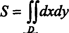
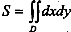
[0017] The orbit is a roughly conical three-dimensional geometric shape. The area was calculated with the orbital ostium as the base of the cone, the vertical distance from the orbital top to the orbital ostium as the cone height, and the volume of the cone as the orbital volume. The orbital volumes of the affected and normal orbits were calculated separately. The orbital volume of the deformed orbit on the affected side is V1, and the normal orbital volume is V0; V1 and V0 are superimposed graphics; the overlapping part is the three-dimensional orbital structure to be reconstructed. For patients with bilateral orbital volume abnormalities (Crouzon syndrome), the orbital anatomy data of normal people were used as reference indicators.

[0018] 2. Research plan

[0019] (1) Computer processing method:

[0020] According to the ...


PUM


Abstract

The invention is particularly suitable to restitution of complex deformity of skull surface. Based on computer image processing and CAD, emulation of 3D segmentation and reconstruction is carried out for structure of skull surface matched to real body. Qualitative even accurate quantitative analyses are carried out for shape and structure of injured skull (including eye socket). The invention discloses layered osteotomy method of splitting off skull socket and surface along natural curvature in middle. The method includes following steps: in operation, fixing moved position promptly; embedding bone tractor to move position gradually; reconstructing skull surface such as inverted valve with vessel pedicle behind the ear. The invention reconstructs fine figure of face to recover normal function.

Description

Technical field [0001] The invention relates to an image-superimposed layered osteotomy method, which relates to the field of medicine, and is especially suitable for the restoration of complicated craniofacial deformities in orthopedic surgery in the field of medicine. Background technique [0002] Congenital craniofacial deformities (craniosynostosis, orbital distance widening, microorbital disease) or craniofacial dysplasia after radiotherapy for juvenile tumors, secondary deformities after trauma, etc., can cause abnormal craniofacial skeleton structure and facial contour Deformation, forming cranio-orbital asymmetry, microorbital disease, severe craniofacial collapse, etc. How to restore the craniofacial appearance to normal is an extremely difficult clinical problem, and some disciplines regard it as a forbidden area. In the late 1970s, with the rise and development of craniofacial surgery in the world, surgical treatment of deformities in the cranium, orbit, face, an...

Claims


Application Information

Patent Timeline
Patent Type & Authority Applications(China)
IPC IPC(8): G06K9/34G06T7/00G06T17/00
Inventor 穆雄铮杨杰
Owner 上海第二医科大学附属第九人民医院
Learn More