Real-time imaging optical coherent endoscope system

An optical coherent, real-time imaging technology, applied in surgery and other fields, can solve the problems of insufficient commercial use, and achieve the effects of simplified data collection and processing, simple structure, good linearity and repeatability

Active Publication Date: 2005-12-21
TSINGHUA UNIV
View PDF2 Cites 16 Cited by
  • Summary
  • Abstract
  • Description
  • Claims
  • Application Information

AI Technical Summary

Problems solved by technology

Although the volume is suitable for blood vessel detection, it is not enough for commercial use because the meshing of gears and the optical coupling efficiency between optical fibers are sensitive to position.

Method used


Image

  • Real-time imaging optical coherent endoscope system
  • Real-time imaging optical coherent endoscope system
  • Real-time imaging optical coherent endoscope system

Examples


Embodiment Construction

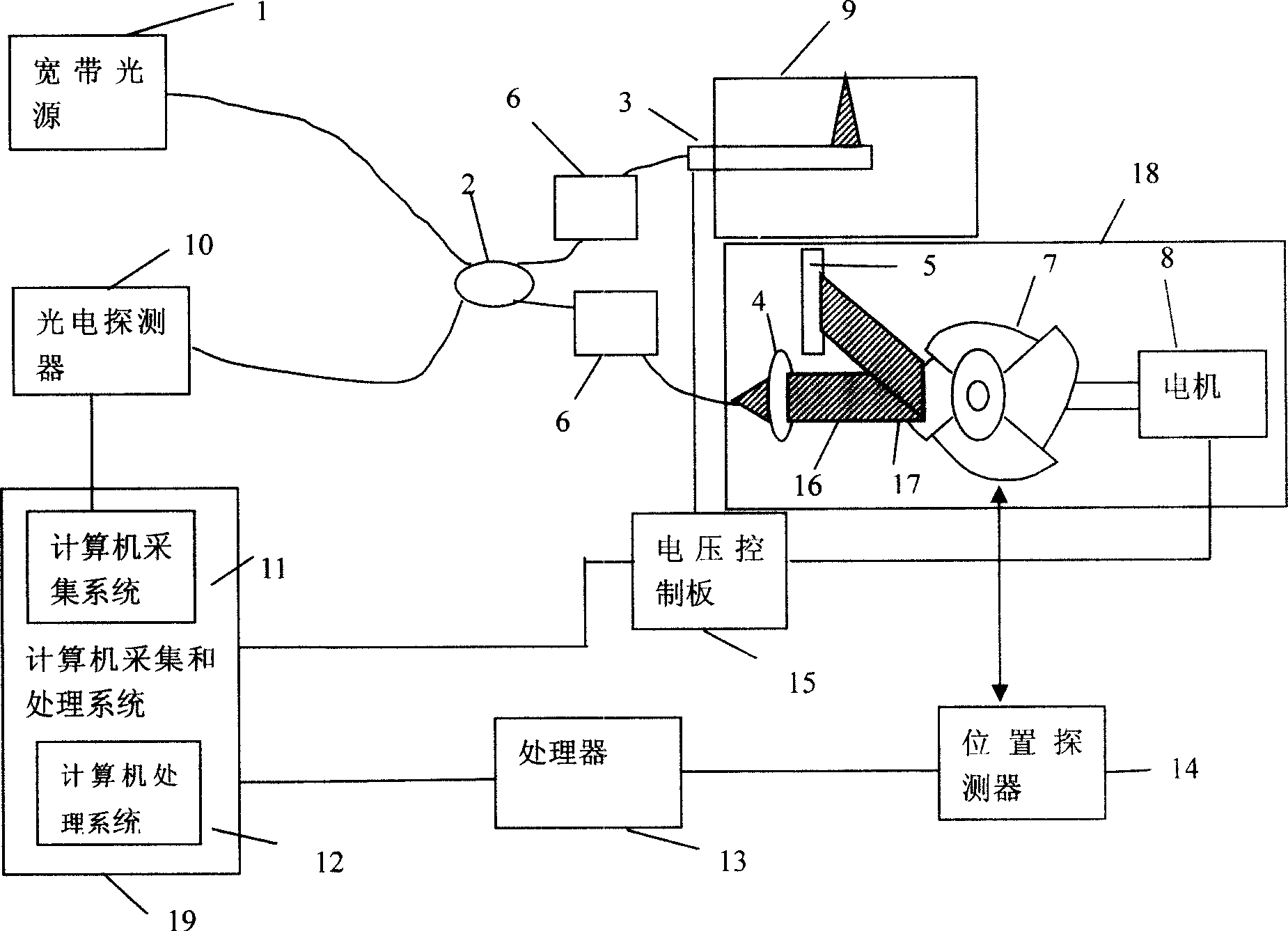
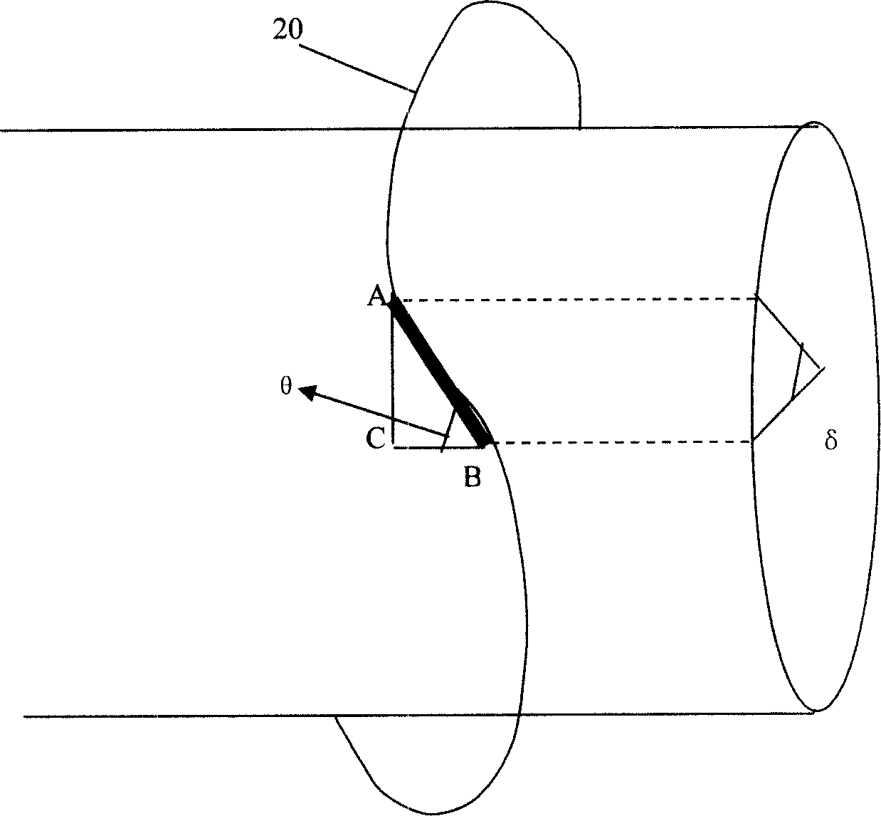
[0017] figure 1 A schematic diagram of the entire imaging system is given. The system comprises a broadband light source 1, an optical fiber coupler 2, a sample arm 3 and a reference arm 18, a photodetector 10 and a computer acquisition and processing system 19, and the reference arm adopts a scanning turntable 7 connected with a motor 8, and the scanning turntable It consists of a central shaft 21 and at least one fan blade 20 whose surface shape is an equidistant helical surface arranged on the central shaft. The broadband light source 1 can use SLD, fiber laser and other light sources, and the central wavelength of the biological tissue light source should be selected around 1.0um. The broadband light source 1 is coupled into the optical fiber through the fiber coupler 2 , and then one is led to the sample arm 3 and the other is led to the reference arm 18 . The light is incident on the fan blade 20 of the scanning turntable through the collimator 4, and is reflected by t...


PUM


Abstract

The present invention relates to a real-time imaged optical coherence endoscopic system. Said invention includes broad-band light source, optical fibre coupler, sample arm and reference arm, position detector and computer collecting and processing system, and said system structure is based on Michelson interferometer, the described reference arm adopts the scanning disk connected with motor, and said scanning disk is formed from central shaft and flabellum which is placed on the central shaft and whose at least one surface form is equidistant spiral surface. The sample arm adopts micro-type optical transmission and scanning device based on ultrasonic motor. Said invention is simple in structure and can provide scanning range greater than 3 mm.

Description

technical field [0001] The invention relates to a technology for performing tomographic imaging by using optical coherence, in particular to an optical coherence endoscopic system for real-time imaging, which belongs to the technical field of medical optical imaging. Background technique [0002] In the last few decades, biological imaging technology has developed rapidly. MRI, XCT, ultrasound, etc. have been widely used in biomedicine. However, these technologies are far from sufficient in applications requiring high resolution (~1um) such as biological slices and tissue analysis. Coronary artery disease is a common disease in modern times, and its early prevention requires high-resolution examination of tissues. OCT is an imaging technology that has been developed in the past ten years. It has many advantages: it can perform micron-resolution cross-sectional imaging of organisms; it can be imaged in real time; It is easy to combine with existing medical devices, so it i...

Claims


Application Information

Patent Timeline
Patent Type & Authority Applications(China)
IPC IPC(8): A61B1/00
Inventor 薛平苗勤
Owner TSINGHUA UNIV
Why Eureka
Try Eureka