
Microelectrode for recording laboratory animal visual sense electrophysiological reaction

A technology of micro-electrodes and experimental animals, which is applied in applications, eye testing equipment, medical science, etc., can solve problems such as easy drift, difficult fixation, large deviation of measurement results, etc., and achieve stable output signals, good repeatability, and record waveforms stable effect

Inactive Publication Date: 2007-02-07
SHANGHAI FIRST PEOPLES HOSPITAL
View PDF0 Cites 1 Cited by
  • Summary
  • Abstract
  • Description
  • Claims
  • Application Information

AI Technical Summary

Problems solved by technology

Because the linear electrode is inconvenient to place and fix in the animal's eye, it is easy to drift and interfere with the recording, so that the recorded waveform has poor repeatability and small amplitude, which directly affects the wide application of visual electrophysiology in experimental animals.
[0009] At the same time, due to the different size and curvature of the cornea of ​​the animal eye, the different eyelids, the shape of the recording electrode and the difference in the manufacturing process, it will greatly affect the recording results.
[0010] In addition, there has been no special reference electrode product suitable for animal visual electrophysiological testing. In actual experiments, a human reference electrode is usually used as a temporary replacement. Since the human reference electrode is a sucker-type electrode, it has more For small rodent experimental animals with hair (such as experimental mice or rabbits, etc.), its fixation is more difficult, and because it cannot be in close contact with the animal's skin, it will bring problems such as inconvenient fixation, poor contact, contact Problems such as large resistance and large deviation of measurement results also lead to poor repeatability and small amplitude of recorded waveforms, which also directly affect the wide application of visual electrophysiology in experimental animals

Method used

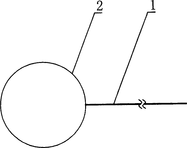
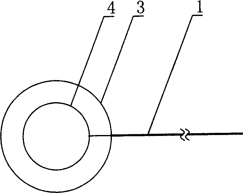
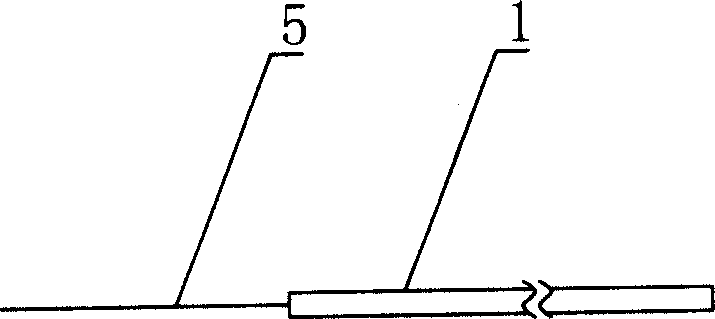

Image

  • Microelectrode for recording laboratory animal visual sense electrophysiological reaction
  • Microelectrode for recording laboratory animal visual sense electrophysiological reaction
  • Microelectrode for recording laboratory animal visual sense electrophysiological reaction

Examples


Embodiment 1

[0043] The recording electrode is a ring-shaped corneal contact electrode, the reference electrode and the ground electrode are needle electrodes, and the experimental animal is a rabbit.

[0044] Drop methylcellulose (or ofloxacin eye ointment) on the corneal surface of rabbit eyes, place the internal / external electrodes with double-circle contact electrodes with diameters of 3mm / 5mm respectively after the corneal surface is anesthetized, and use needles for reference electrodes and ground electrodes. Electrodes were inserted into the ipsilateral ear and the top of the head, respectively.

[0045] Using the above-mentioned miniature electrodes, in cooperation with the LKC UTAS-2000 electrophysiological instrument in the United States, the dual-channel ERG is recorded, and the recorded waveform is as follows: Figure 4 shown.

[0046] It can be seen from the figure that there is basically no interference clutter (that is, no steep peak) in the a→b segment of the displayed wav...

Embodiment 2

[0048] The recording electrode is a ring-shaped corneal contact electrode, the reference electrode and the ground electrode are needle electrodes, and the experimental animals are BN (Brown Norway) rats.

[0049] The recording electrode adopts a 0.8mm / 1.5mm double-circle contact electrode, which is placed directly into the eyeball after the animal is anesthetized. The surface is dripped with physiological saline or methylcellulose, and the reference electrode and the ground electrode are needle electrodes, which are respectively inserted into the same side of the ear. and top of head.

[0050] Using the above-mentioned miniature electrodes, in cooperation with the LKC UTAS-2000 electrophysiological instrument in the United States, the dual-channel ERG is recorded, and the recorded waveform is as follows: Figure 5 shown.

[0051] It can be seen from the figure that the segment N1→O4 in the displayed waveform curve has no interference clutter, and the waveform curve is relativ...

Embodiment 3

[0054] The recording electrode, the reference electrode and the grounding electrode are metal needle-shaped electrodes with a 1ml stainless steel needle for injection as the needle tip, and the experimental animals are Wistar rats.

[0055] The recording electrode was inserted under the skin connecting the two ears, and the reference electrode and the grounding electrode were inserted into the ipsilateral ear and the top of the head respectively.

[0056] Get the FVEP record waveform as Image 6 shown.

[0057] It can be seen from the figure that the displayed waveform curve has no clutter interference, and the waveform is stable, reflecting the advantages of the technical solution, which is convenient and accurate in positioning, easy to fix, good in contact, stable in recorded waveform, and good in repeatability of the recorded waveform.

[0058] All the other are with embodiment 1 or 2.


PUM


Abstract

The invention discloses a minimized electrode in the physiological checking element domain, which consists of recording electrode and or reference electrode, wherein the recording electrode is composed of conductive electrode rod and electrode on one end of conductive rod; the electrode is ring-shaped metal corneal contacting electrode; or the recording electrode and or reference electrode is composed of insulating conductive electrode rod and electrode on one end of rod conduct; the electrode is needle-shaped metal electrode.

Description

technical field [0001] The invention belongs to the field of physiological test instruments, in particular to a device for measuring animal bioelectric signals. Background technique [0002] Visual electrophysiological technology is the only objective visual function testing method commonly used in ophthalmology clinics. Since its clinical application for more than half a century, its emergence and development are inseparable from the basis of animal experiments. [0003] The non-invasiveness and objectivity of visual electrophysiological technology, the ability to go deep into the molecular level under special conditions, and the hierarchical positioning and accuracy of detection of retinal and visual path lesions make this technology widely used in clinical ophthalmology and even in neurology departments. It plays a very important and irreplaceable role in related cross research. [0004] In the past 10 years, visual electrophysiological technology has advanced by leaps a...

Claims


Application Information

Patent Timeline
Patent Type & Authority Applications(China)
IPC IPC(8): A61B3/10
Inventor 吴晓平张皙宫媛媛吴星伟许迅
Owner SHANGHAI FIRST PEOPLES HOSPITAL
Learn More