Subdural electrode localization and visualization using parcellated, manipulable cerebral mesh models

a cerebral mesh model and subdural electrode technology, applied in tomography, instruments, applications, etc., can solve the problems of significant problems adversely affecting the accurate localization of electrodes located underneath the craniotomy, so as to achieve optimal electrical signal recording, accurate electrode placement, and correct identification of sites

Active Publication Date: 2018-12-11
BOARD OF RGT THE UNIV OF TEXAS SYST
View PDF26 Cites 14 Cited by
  • Summary
  • Abstract
  • Description
  • Claims
  • Application Information

AI Technical Summary

Benefits of technology

[0009]The present invention relates to methods for localization and visualization of implanted electrodes, such as, for example, subdural electrodes (SDEs) and depth electrodes (DEs), particularly to localizing and visualizing implanted electrodes to the cortical surface and / or particular volumes of a patient's brain, and more particularly to methods of accurately mapping implanted electrodes to the cortical topology and / or associated topological volumes of a patient's brain. This invention further relates to methods of surgical intervention utilizing accurate cortical surface modeling and / or topological volume modeling of a patient's brain for placement of electrodes and / or utilization thereof for surgical intervention, such as for accurately placing electrodes for optimal recording of electrical signals and / or properly identifying sites for surgical intervention.
[0012]In general as disclosed herein, precise localization as well as optimized viewing of implanted intracranial electrodes may be shown on a manipulable 3D cortical mesh model that may be parcellated, such as by automatic manipulation, to show anatomic details of specific brain regions (e.g. specific cortical gyri, functional zones), such as where the electrodes are placed. Thus, both precise localization as well as optimized viewing of implanted intracranial electrodes may be accomplished. For example, the mesh model may be morphed into unnatural shapes, such as, for example, a dilated, smoothed pial surface, that may allow for viewing the areas that individual electrodes are in contact with, as well as the regions where such recording / stimulation is not happening, thus providing a holistic view of the data. Further, the localization of electrodes onto an individually parcellated cortical surface may allow for the a priori assignment of putative functional zones relative to each electrode. This may, for example, augment, verify or altogether replace imaging based functional mapping techniques (e.g. MEG or fMRI) in some cases, and / or electrical cortical stimulation mapping in others.
[0017]In some exemplary embodiments, the visual registration of a set of implanted electrodes may utilize pre-implantation and post-implantation imaging of the brain in an electrode array implantation, such as by utilizing digital and / or analog photography of the operative site in an OR. Pre-implantation imaging may generally provide visual information of the underlying cortical surface prior to implantation of the electrodes and may thus be utilized to compare anatomical features with the 3D model, such as to establish anatomical landmarks. After implantation of the electrodes, post-implantation imaging may be performed to visualize the electrodes and the anatomical features of the brain. In one embodiment, a skilled practitioner, such as, for example, a physician or veterinarian, may readily compare the anatomical features from the intraoperative imaging to the 3D model, such as by matching and / or aligning anatomical landmarks, to visually translate the locations of the electrodes from the intraoperative imaging to the 3D model.
[0022]In still another aspect of the present invention, electrical signals from implanted electrodes may be mapped to the 3D cortical surface to more accurately determine the particular region(s) of the patient's brain contributing to a particular signal at an electrode. These electrical signals may reflect normal ongoing brain activity, activity stimulated by certain normal behaviors (e.g. movement, language) or pathological activity such as seen in epilepsy. In general, traditional methods typically map electrical activity at an electrode to regions of the brain that are closest in Euclidian distance to the electrode which, without being bound to any particular theory, may erroneously attribute electrical activity to topologically distant portions of the brain due to the Euclidian distance not accounting for surrounding topology about the electrode.

Problems solved by technology

Various physiological issues, such as, for example, blood and fluid accumulation underneath the craniotomy flap, may lead to a non-linear deformation of the brain surface and of an electrode array on post-operative CT scans, and may adversely impact the accurate localization of electrodes located underneath the craniotomy.
Prior methods that localize electrodes based on their identification on a post-implantation CT, with co-registration to a pre-implant MRI may further result in significant problems with accuracy of the electrode localization.

Method used


Image

  • Subdural electrode localization and visualization using parcellated, manipulable cerebral mesh models
  • Subdural electrode localization and visualization using parcellated, manipulable cerebral mesh models
  • Subdural electrode localization and visualization using parcellated, manipulable cerebral mesh models

Examples


Embodiment Construction

[0045]The detailed description set forth below is intended as a description of the presently exemplified methods, devices and compositions provided in accordance with aspects of the present invention, and is not intended to represent the only forms in which the present invention may be practiced or utilized. It is to be understood, however, that the same or equivalent functions and components may be accomplished by different embodiments also intended to be encompassed within the spirit and scope of the invention.

[0046]Unless defined otherwise, all technical and scientific terms used herein have the same meaning as commonly understood to one of ordinary skill in the art to which this invention belongs. Although any methods, devices and materials similar or equivalent to those described herein can be used in the practice or testing of the invention, the exemplified methods, devices and materials are now described.

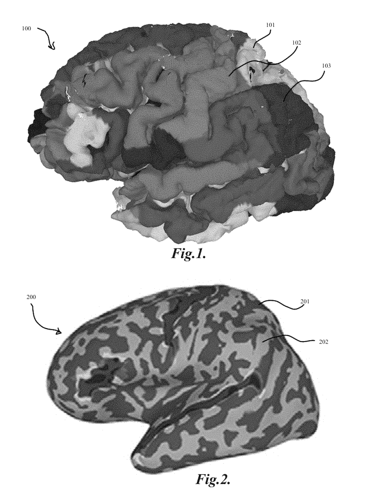
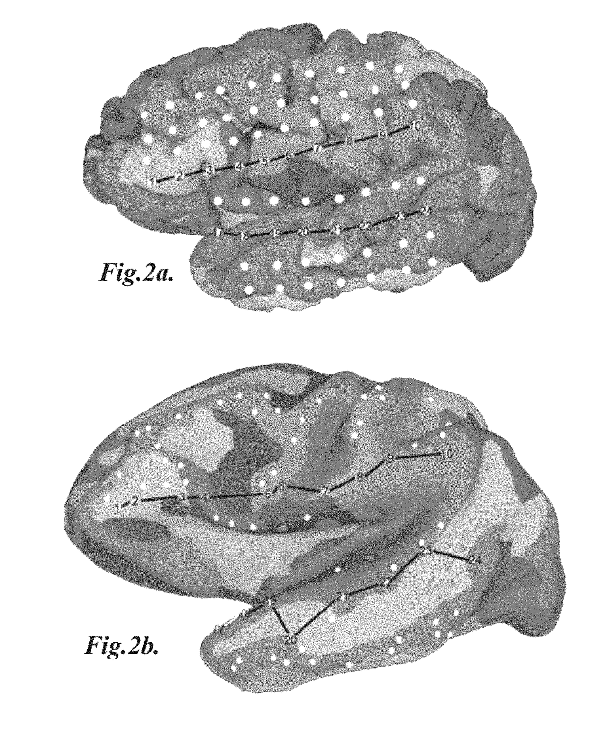
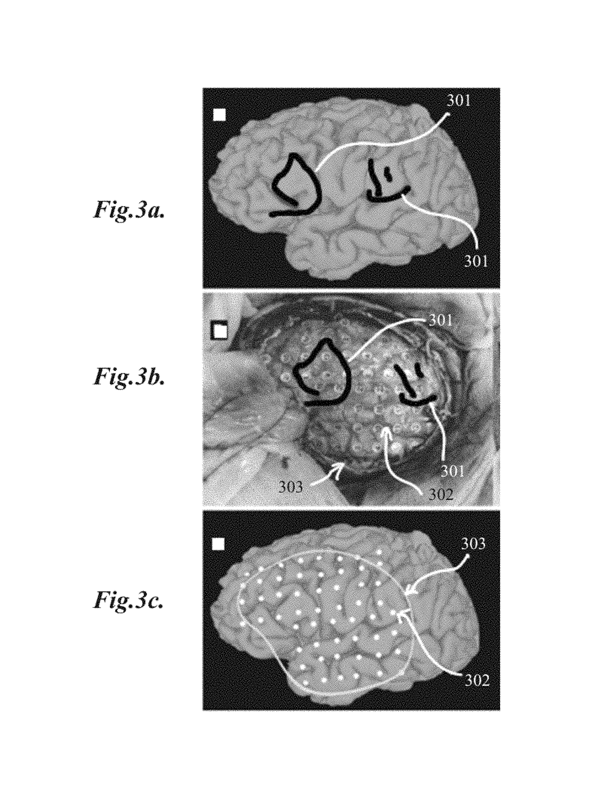
[0047]The present invention relates to methods for localization and visu...


PUM


Abstract

This invention relates generally to methods for localization and visualization of implanted electrodes and penetrating probes in the brain in 3D space with consideration of functional brain anatomy. Particularly, this invention relates to precise and sophisticated methods of localizing and visualizing implanted electrodes to the cortical surface and / or topological volumes of a patient's brain using 3D modeling, and more particularly to methods of accurately mapping implanted electrodes to the cortical topology and / or associated topological volumes of a patient's brain, such as, for example, by utilizing recursive grid partitioning on a manipulable virtual replicate of a patient's brain. This invention further relates to methods of surgical intervention utilizing accurate cortical surface modeling and / or topological volume modeling of a patient's brain for targeted placement of electrodes and / or utilization thereof for surgical intervention in the placement of catheters or other probes into it.

Description

CROSS-REFERENCE TO RELATED APPLICATIONS[0001]This application claims the benefit and priority of U.S. provisional patent application Ser. No. 61 / 951,861, filed Mar. 12, 2014, entitled “Subdural Electrode Localization and Visualization Using Parcellated, Manipulable Cerebral Mesh Models”, the contents of which is hereby incorporated by reference in its entirety.STATEMENT UNDER 35 U.S.C. § 202(C)(6)[0002]These inventions were made with U.S. Government support under Grant Nos. RR024149 and RR024148 awarded by the National Institutes of Health. The government has certain rights in the invention.COPYRIGHT NOTICE[0003]A portion of the disclosure of this patent document contains material that is subject to copyright protection. The copyright owner has no objection to the facsimile reproduction by anyone of the patent document or the patent disclosure, as it appears in the U.S. Patent and Trademark Office patent file or records, but otherwise reserves all copyright rights whatsoever.FIELD O...

Claims


Application Information

Patent Timeline
Patent Type & Authority Patents(United States)
IPC IPC(8): A61B5/05A61B5/00A61B5/055A61B6/03A61B6/00A61B5/0476
CPCA61B5/0042A61B5/055A61B5/0037A61B5/0077A61B6/032A61B6/5205A61B6/4417A61B5/0476A61B5/061A61B6/12A61B6/501A61B6/5247A61B5/24A61B5/369A61B2034/105A61B2034/2055A61B5/37G06T15/04A61B2576/026A61B5/384
Inventor TANDON, NITINCONNER, CHRISTOPHER R.PIETERS, THOMAS A.KADIPASAOGLU, CIHAN MEHMET
Owner BOARD OF RGT THE UNIV OF TEXAS SYST
Why Eureka
Try Eureka