
MicroRNAs for cardiac regeneration through induction of cardiac myocyte proliferation

a technology of cardiac myocytes and micrornas, which is applied in the field of micrornas for cardiac regeneration through induction of cardiac myocyte proliferation, can solve the problems of compromising cardiac function, poor long-term prognosis of heart failure patients, and very restricted ability of mammalian adult heart to repair itself after injury, etc., and achieves the effect of increasing the number of cardiomyocytes and increasing the proliferation of fully differentiated rat cardiomyocytes

Active Publication Date: 2019-07-02
ROLITH INC +1
View PDF15 Cites 1 Cited by
  • Summary
  • Abstract
  • Description
  • Claims
  • Application Information

AI Technical Summary

Benefits of technology

The selected microRNAs significantly increase cardiomyocyte proliferation in vitro and in vivo, improving heart function and cardiac tissue regeneration, offering a novel approach for treating heart diseases like myocardial infarction, cardiomyopathy, and heart failure.

Problems solved by technology

Despite pharmacological treatment, long-term prognosis of heart failure patients remains poor: more than 60% of these patients die within 5 years, from a worsening of the disease or due to sudden ventricular arrhythmias.
As a consequence of this limited proliferation capacity of adult cardiomyocytes, the ability of the mammalian adult heart to repair itself following injury is very restricted (Senyo et al.
This significantly compromises cardiac function, which often tends to worsen over time, leading to heart failure.
However, the exact molecular mechanisms controlling this transition and those responsible for sustaining proliferation and differentiation of cardiac myocytes remain largely unknown.
To date, no exhaustive search for microRNAs able to induce cardiac regeneration has been performed yet.
However, there is no explicit disclosure of microRNAs that were specifically selected for their capacity of stimulating the proliferation of adult cardiomyocytes in the cardiac tissue which is damaged or impaired in its physiological function, thus permitting cardiac regeneration in vivo.
This person is also aware that the therapy for the treatment of acute phase is unsuitable for the treatment of heart diseases where cardiomyocyte proliferation is necessary for heart regeneration.

Method used


Image

  • MicroRNAs for cardiac regeneration through induction of cardiac myocyte proliferation
  • MicroRNAs for cardiac regeneration through induction of cardiac myocyte proliferation
  • MicroRNAs for cardiac regeneration through induction of cardiac myocyte proliferation

Examples


example 1

Functional Screening Identifies MicroRNAs that Control Proliferation of Rat Neonatal Cardiomyocytes In Vitro

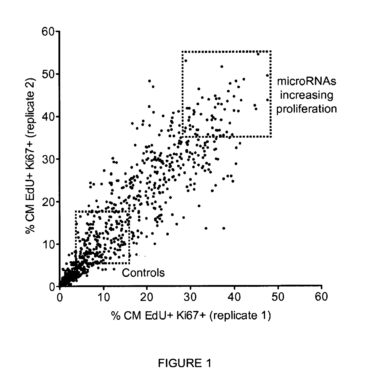
[0115]Given the involvement of microRNAs in the regulation of several biological processes, including cell proliferation, we wanted to investigate whether microRNAs might control proliferation of primary cardiac myocytes ex vivo and identify the microRNAs most effective in increasing the proliferative capacity of these cells.

[0116]To tackle this issue, we performed a fluorescence microscopy-based high-throughput screening in rat neonatal cardiomyocytes using a commercial library of 988 microRNA mimics (miRIDIAN microRNA mimics, Dharmacon, Thermo Scientific) corresponding to all the annotated human microRNAs (according to miRBase release 13.0, 2009). Cells were stained with the nuclear dye Hoechst 33432, antibodies against the cardiomyocyte marker sarcomeric alpha-actinin and the proliferation antigen Ki-67, and with EdU, an uridine analogue that is incorporated into newly synt...

example 2

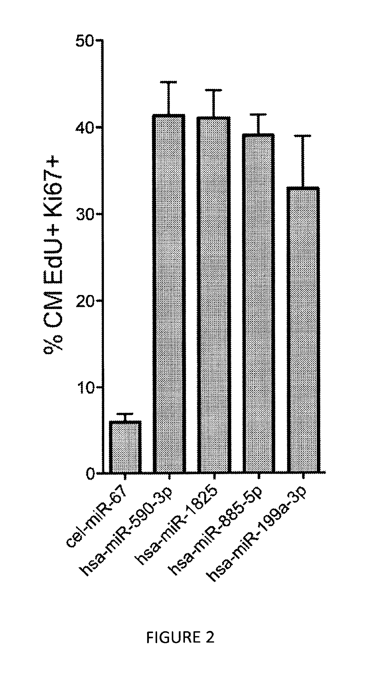
A Subset of MmicroRNAs Control In Vitro Proliferation of Cardiomyocytes from Different Organisms (Rat, Mouse and Human)

[0125]During development, cardiomyocytes in the mouse heart stop dividing sooner than their counterparts in the rat heart (shortly after birth and 3 to 4 days after birth, respectively). As a consequence of this, cardiomyocytes isolated from newborn mice have a proliferative capacity significantly lower than those isolated from rats of the same age; while proliferation of cardiomyocytes isolated from neonatal rats (post-natal day 0) is ca. 12.5%, that of mice cardiomyocytes is ca. 5%.

[0126]We therefore repeated the fluorescence microscopy-based high-throughput experiments described in Example 1 in mouse neonatal cardiomyocytes, using the 208 selected microRNAs that were shown to increase proliferation of rat cardiomyocytes. Results from these experiments showed that out of the 208 microRNAs tested, 36 microRNAs also increased the proliferation of mouse cardiomyocyte...

example 3

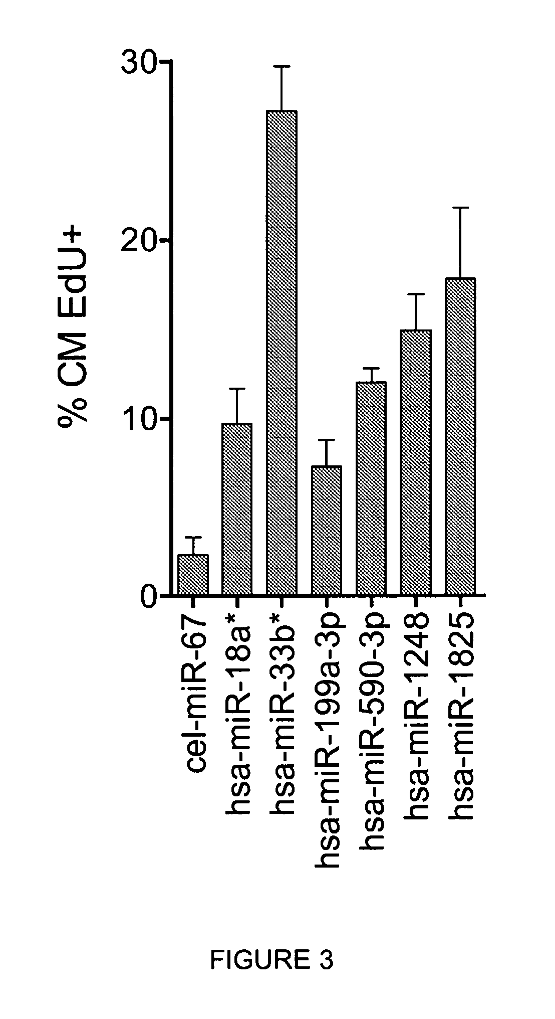
MicroRNAs Increase Cardiac Myocytes Mitosis, Cell Division (Cytokinesis) and Cell Number In Vitro

[0133]To demonstrate that the increase in cardiomyocyte proliferation, as assessed by staining with Ki-67 proliferation marker and EdU incorporation during DNA synthesis, correlates with an increase in cardiomyocyte cell division events (cytokinesis), cardiomyocytes were treated with individual microRNAs and additional markers were evaluated, namely: i) staining for histone H3 phosphorylated at Serine 10(P-S10-H3), which detects cells in late G2 / mitosis; ii) staining for AuroraB kinase, a component of midbodies, a transient structure which appears near the end of cytokinesis just prior to, and is maintained for a brief period after, the complete separation of the dividing cells; and iii) cardiomyocyte number at 6 days after microRNA transfection.

[0134]Treatment of cardiomyocytes with the selected microRNAs led to a significant increase in both the number of cells positive for P-S10-H3 (f...


PUM

PropertyMeasurementUnit
timeaaaaaaaaaa
total volumeaaaaaaaaaa
sizeaaaaaaaaaa

Abstract

The present invention discloses a set of human microRNAs, or a primary transcript for such microRNAs, or a precursor of such microRNAs, or a mimic of such microRNAs or a combination thereof, and their use as medicaments for inducing proliferation of cardiomyocytes for the prevention and treatment of heart diseases associated with a loss of cardiomyocytes. The invention also relates to a method for screening microRNAs and biological and therapeutically active compounds for their ability to increase proliferation of cardiomyocytes.

Description

[0001]This is the U.S. national stage of Application No. PCT / IB2012 / 057590, filed Dec. 21, 2012; which claims priority benefit of Italian Application No. RM2011A000685, filed Dec. 23, 2011; the entire contents of each of which are incorporated by reference herein.[0002]The present invention relates to the fields of pharmaceuticals and biotechnology. In particular, the present invention discloses a set of human microRNAs and their use as medicaments, especially as gene therapy, to induce proliferation of cardiomyocytes as a mean to stimulate cardiac regeneration for the treatment of heart diseases associated with a loss of cardiomyocytes (myocardial infarction, cardiomyopathy of ischemic and non-ischemic origin, myocarditis and heart failure).[0003]In addition, the invention relates to a method for screening of biological and pharmaceutical active compounds for their ability to increase proliferation of cardiomyocytes.BACKGROUND OF THE INVENTION[0004]Despite the enormous progress mad...

Claims


Application Information

Patent Timeline
Patent Type & Authority Patents(United States)
IPC IPC(8): C12N15/11G01N33/50C12N5/077C12N15/113A61K48/00
CPCC12N15/113C12N5/0657G01N33/5061A61K48/00G01N2500/10C12N2320/30C12N2501/65C12N2506/02C12N2310/141A61P43/00A61P9/00A61P9/04A61P9/10
Inventor GIACCA, MAUROZACCHIGNA, SERENACUNHA MANO, MIGUEL LUISBREGIEIRO EULALIO, ANA SOFIA
Owner ROLITH INC
Learn More