

Methods and test kits for determining male fertility status
a technology for determining male fertility and testing kits, applied in the field of male fertility, can solve the problems of not being able to immediately fertilize an egg, and achieve the effects of preventing shearing of the sperm membrane, avoiding damage to the sperm membrane, and sufficient siz
- Summary
- Abstract
- Description
- Claims
- Application Information
AI Technical Summary
Benefits of technology
Problems solved by technology
Method used
Image

Examples
example 1
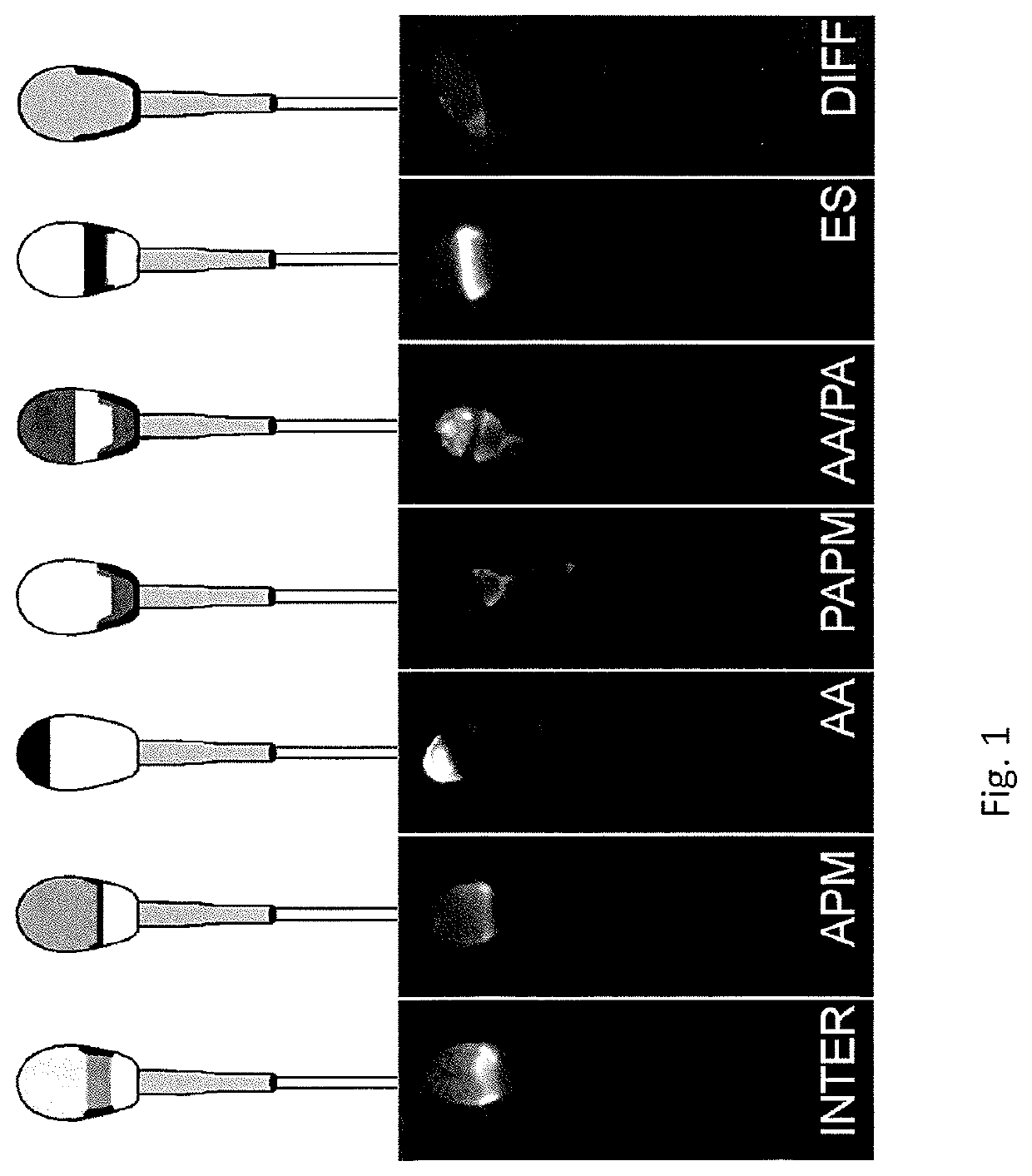
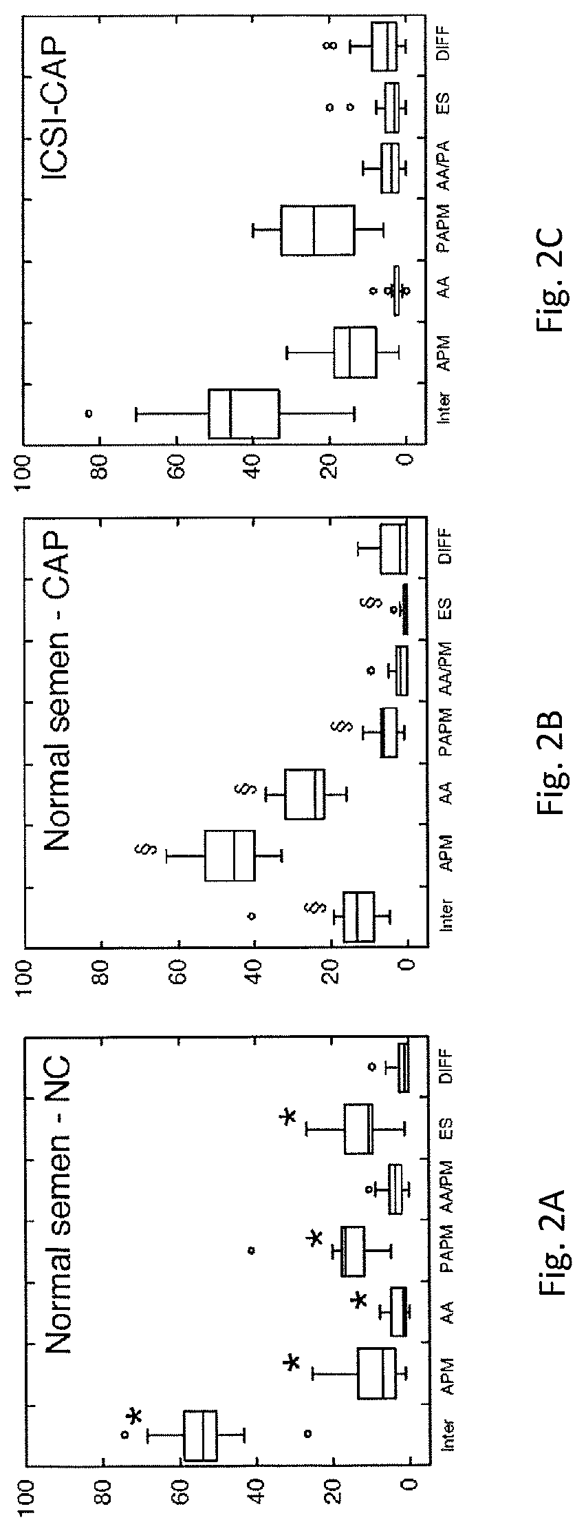
[0137]This example provides demonstration of GM1 localization patterns obtained with human sperm. Ejaculated sperm were collected from male donors, and allowed to liquefy for 20 mins at 37° C., and then volume, initial count, motility and morphology assessments were performed. 1 ml of the semen sample was layered on top of 1 ml of a density gradient (90% Enhance-S; Vitrolife, San Diego, Calif., USA) in a 15 ml conical tube. The tube was centrifuged at 300×g for 10 minutes. The bottom 1 ml fraction was transferred to a new 15 ml tube and then resuspended in 4 ml of mHTF. This was centrifuged at 600×g for 10 minutes. The supernatant was removed and the pellet of sperm was resuspended in 0.5 ml of mHTF. The washed sperm were then evaluated for concentration and motility. Equal volumes of sperm were then added to two tubes, such that the final volume of each tube was 300 μl, and the final concentration of sperm was 1,000,000 / ml. The first tube contained mHTF (non-capacitating condition)...
example 2
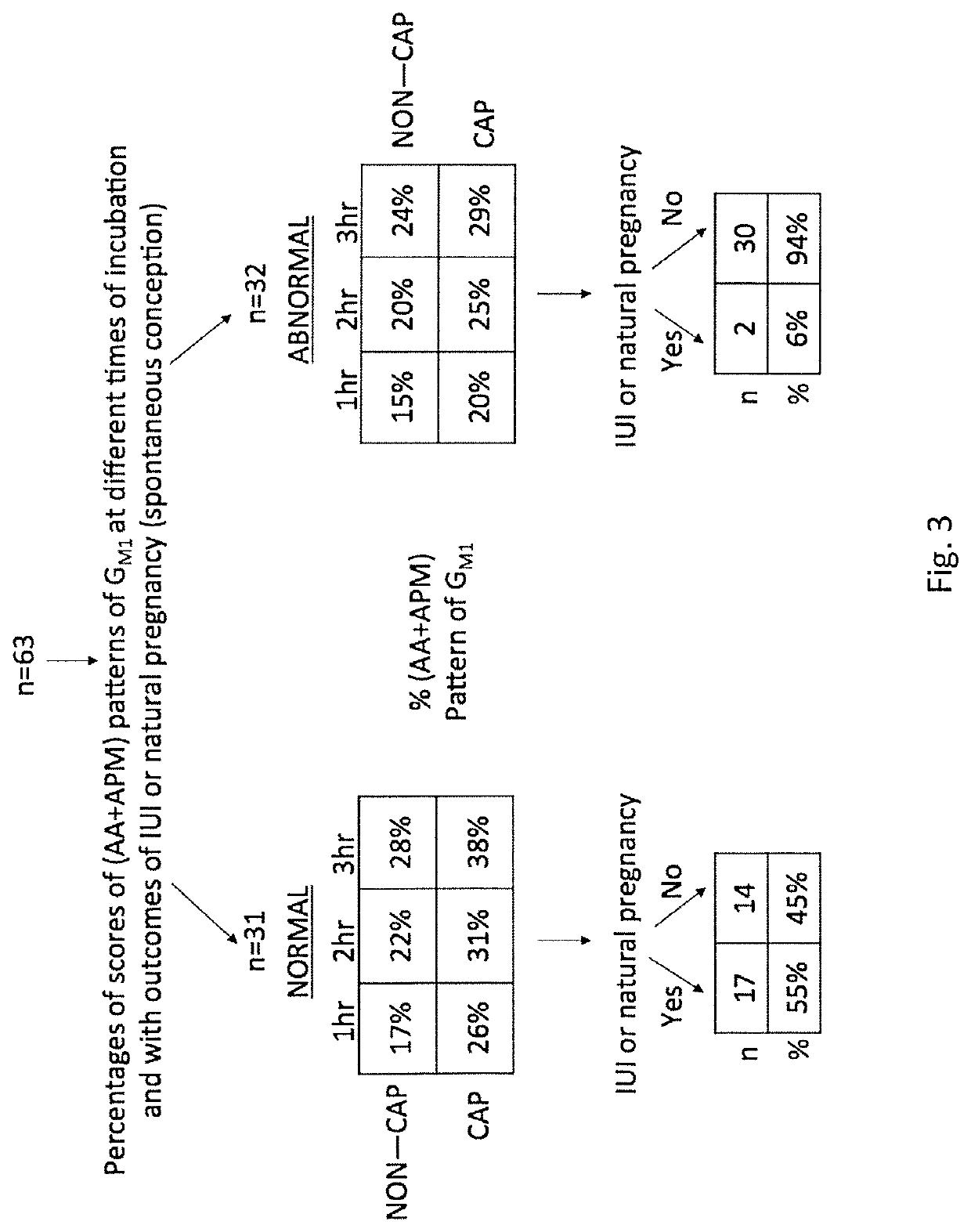
[0140]In this example, clinical histories of 34 patients were studied to perform a close analysis of their GM1 assay scores relative to history of ever achieving clinical evidence of pregnancy. A male patient was defined as “fertile” if a patient couple achieved some evidence of fertilization / clinical pregnancy (even if limited to biochemical evidence or a sac without heartbeat on ultrasound) within 3 or fewer cycles.
[0141]Analysis of the data for these 34 patients revealed that if one applied a cut-off of 40% (APM+AA) for the score of the capacitated samples at the 3 hour time point, then ⅞ who “passed” (having a score of 39.5% or greater), were found to have been designated “fertile” (87.5%). Of the 26 who “failed” (having a score of 39.4 or less), only 3 / 26 had evidence of clinical pregnancy (11.5%). (see Table 1 below).
[0142]If one reduces the cutoff, it would be predicted that more people who are clinically sub-fertile will get a passing score and the percentage that pass the a...
example 3
[0147]Sperm cells were treated as described in Example 1 but incubated in fixative for 24 hours. The labeled sperm cells were then evaluated by fluorescence microscopy. A new GM1 localization pattern, Lined-Cells was identified as illustrated in FIGS. 6A, 6B, 6C and 6D. In Lined-cells, as illustrated in FIG. 6A, there is GM1 signal at the bottom of the equatorial segment / top of the post acrosomal region, and at the plasma membrane overlying the acrosome. The signal is evenly distributed in the post acrosome / equatorial region and the plasma membrane overlying the acrosome. There is also a band at the equatorial segment that lacks signal. As illustrate in FIG. 6B, the signal at the plasma membrane overlying the acrosome is brighter than the signal at the post acrosome / equatorial band. As illustrated in FIGS. 6C and 6D, the signal found at the post acrosome / equatorial band is brighter than the signal at the plasma membrane overlying the acrosome.
[0148]Sperm cells from a single donor we...
PUM
| Property | Measurement | Unit |
|---|---|---|
| time period | aaaaa | aaaaa |
| time period | aaaaa | aaaaa |
| time period | aaaaa | aaaaa |
Abstract
Description
Claims
Application Information