Camera positioning system, method, and apparatus for capturing images during a medical procedure

a positioning system and camera technology, applied in the field of imaging camera positioning system, can solve the problems of poor resolution, large size, and the lack of known camera system, and achieve the effect of convenient insertion

Active Publication Date: 2020-04-21
TYCO HEALTHCARE GRP LP
View PDF21 Cites 0 Cited by
  • Summary
  • Abstract
  • Description
  • Claims
  • Application Information

AI Technical Summary

Benefits of technology

[0005]The visualization device of any of preceding paragraphs and / or any of visualization devices described below can include one or more of the following features. The device can include a plurality of openings positioned on the exterior of the housing and configured to removably receive a plurality of supporting pins attached to the mounting interface of the robotic surgery apparatus. Rotation of the plurality of rollers can cause a diameter of the loop formed by the camera tube to change. Rotation of the plurality of rollers in the first direction can cause the diameter of the loop to decrease and rotation of the plurality of rollers in the second direction can cause the diameter of the loop to increase.
[0008]The visualization device of any of preceding paragraphs and / or any of visualization devices described below can include one or more of the following features. At least one camera can include two cameras configured to provide a stereoscopic image of a region of interest in the body cavity, and the device further can include a light source configured to illuminate the region of interest. Plurality of rollers can include surfaces with a friction coefficient that allows the plurality of rollers to slip along the portion of the camera tube that passes through the drive opening of the housing to prevent movement of the portion of the camera tube. Plurality of rollers can provide a sterile barrier between a sterile camera tube and non-sterile mounting interface of the robotic surgery apparatus when the housing is attached to the mounting interface of the robotic surgery apparatus.
[0012]The visualization device of any of preceding paragraphs and / or any of visualization devices described below can include one or more of the following features. At least one driver can include a plurality of rollers configured to contact the camera tube and advance the camera tube toward the region of interest and retract the camera tube away from the region of interest. Rotation of the plurality of rollers in the first direction can cause the camera tube to advance and a diameter of the loop to decrease. Rotation of the plurality of rollers in the second direction opposite to the first direction can causes the camera tube to retract and diameter of the loop to increase.
[0016]In some cases, an insertion device for a single port robotic surgery apparatus can include a first portion including a plurality of instrument channels positioned in an interior of the first portion and extending along substantially an entire length of the first portion, the plurality of instrument channels configured to removably house a plurality of surgical instruments and first and second camera channels positioned in the interior of the first portion and extending along substantially the entire length of the first portion, the first camera channel configured to removably house a primary camera tube and the second camera channel comprising a secondary camera. The insertion device can also include a second portion including an insertion channel terminating at a first end with a first opening configured to permit the primary camera tube to pass through and terminating at a second end opposite the first end with a second opening aligned with the first camera channel of the first portion, the insertion channel configured to permit the primary camera tube to pass through the insertion channel and enter the first camera channel. The secondary camera facilitates insertion into a body cavity of at least one of the plurality of instruments or the primary camera tube.

Problems solved by technology

Known camera systems suffer from a variety of shortcomings, including large size, poor resolution, obstacles with being sterilized, lack of reliability, difficulties with being replaced during medical procedure, and the like.
Actuating the one or more second actuators can cause one or more flexible links of the primary camera to be manipulated, which can cause articulation of the distal end.

Method used


Image

  • Camera positioning system, method, and apparatus for capturing images during a medical procedure
  • Camera positioning system, method, and apparatus for capturing images during a medical procedure
  • Camera positioning system, method, and apparatus for capturing images during a medical procedure

Examples


Embodiment Construction

Overview

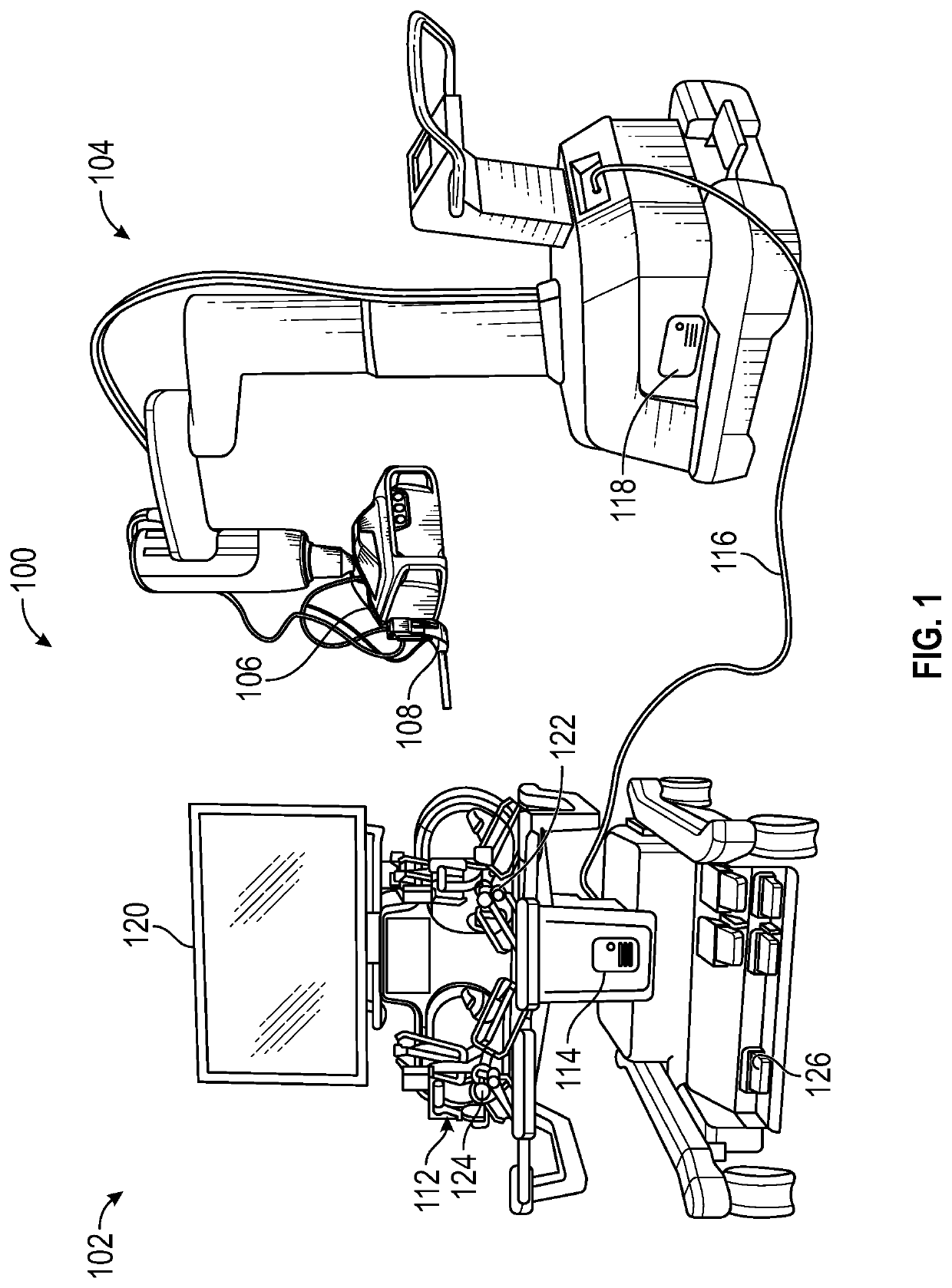
[0044]When performing medical procedures (for example, with assistance of surgery using a robotic surgical system) one or more instruments can be inserted into a body cavity of a patient. The insertion process has some risk since instruments may inadvertently damage organs or tissue while being inserted. Incorrect positioning of the one or more instruments in the body cavity may also result in a limited range of motion within the body cavity.

[0045]As an example, when performing abdominal surgery, at least one incision would be made in a body wall of the patient's abdomen. A trocar or other access port, may then be inserted through the incision. A camera can be first inserted through the access port and used by a surgeon to capture and relay stereoscopic images of a surgical site. One or more instruments can be inserted following the camera insertion. Views provided by the camera facilitate insertion of the one or more instruments and their manipulation of the surgical site.

[...


PUM


Abstract

In some embodiments, an insertion device for a robotic surgery apparatus can include a first portion with a plurality of instrument channels configured to removably house a plurality of surgical instruments and first and second camera channels positioned in an interior of the first portion and extending along substantially the entire length of the first portion. The first camera channel can removably house a primary camera tube, and the second camera channel can include a secondary camera. The device can include a second portion with an insertion channel terminating at a first end with a first opening permitting the primary camera tube to pass through and terminating at a second end opposite the first end with a second opening aligned with the first camera channel of the first portion. The insertion channel can permit the primary camera tube to pass through the insertion channel and enter the first camera channel.

Description

TECHNICAL FIELD[0001]This disclosure relates generally to positioning a camera for imaging and more particularly to positioning a camera inside a body cavity of a patient for capturing images during a medical procedure.DESCRIPTION OF RELATED ART[0002]Miniaturized cameras are used during investigative medical procedures and surgical procedures, such as laparoscopic surgery and computer assisted robotic surgery, to produce images of a site of the procedure within a body cavity of the patient. A camera generally includes an illumination source for illuminating the site of the procedure and a lens for capturing images of the site. Known camera systems suffer from a variety of shortcomings, including large size, poor resolution, obstacles with being sterilized, lack of reliability, difficulties with being replaced during medical procedure, and the like. The present disclosure overcomes these and other problems associated with known camera systems, methods, and apparatuses.SUMMARY[0003]In...

Claims


Application Information

Patent Timeline
Patent Type & Authority Patents(United States)
IPC IPC(8): A61B1/012A61B90/00A61B34/20A61B1/00A61B1/018A61B90/50A61B34/30A61B1/06A61B34/10A61B34/37
CPCA61B1/012A61B1/00135A61B1/0125A61B1/018A61B90/361A61B34/20A61B1/0684A61B34/37A61B90/50A61B2034/107A61B2034/305A61B1/00105A61B34/10A61B2034/301A61B1/05A61B1/00188A61B1/00193A61B1/07A61B2090/371A61B90/37A61B2090/306A61B2090/309A61B2017/00477A61B46/10A61B34/30A61B34/74A61B2034/742
Inventor GENOVA, PERRY A.PFLAUMER, HANS CHRISTIANLAAKSO, AKI HANNU EINARIKATZ, ALLANESPINOSA, ALEJANDROAMPUERO, EDUARDO A.
Owner TYCO HEALTHCARE GRP LP
Why Eureka
Try Eureka