Method of screening for colorectal cancer

a colorectal cancer and neoplasm technology, applied in the field of colorectal cancer classification, can solve the problems of inconvenient diagnosis, inability to accurately predict the risk of cancer, and inability to reliably diagnose colorectal cancer symptoms

Active Publication Date: 2021-03-09
CLINICAL GENOMICS PTY LTD
View PDF3 Cites 1 Cited by
  • Summary
  • Abstract
  • Description
  • Claims
  • Application Information

AI Technical Summary

Benefits of technology

[0082]Without limiting the present invention to any one theory or mode of action, the skilled person may screen one or more subregions for each gene marker.

Problems solved by technology

These mushroom-shaped growths are usually benign, but some develop into cancer over time.
However, this is related to size of an adenoma.
Except for the presence of adenomas and its size, none of these is objectively defined, and all those other than number and size are subject to observer error and to confusion as to precise definition of the feature in question.
Because such factors can be difficult to assess and define, their value as predictors of current or future risk for cancer is imprecise.
Unfortunately, many of the symptoms may occur in other diseases as well, and hence symptoms may not be conclusively diagnostic of colorectal cancer.
A tumour that is large enough to fill the entire lumen of the bowel may cause bowel obstruction.
This occasionally leads to the obstructed and distended bowel perforating and causing peritonitis.
The disease may invade other organs, and may cause blood or air in the urine or vaginal discharge.
Colorectal cancer may also lead to weight loss, generally due to a decreased appetite.
This may go unnoticed, but large deposits in the liver may cause jaundice and abdominal pain (due to stretching of the capsule).
Even modest efforts to implement colorectal cancer screening methods can result in a drop in cancer deaths.
Despite this, colorectal cancer screening rates remain low.
In the context of large intestine neoplasms, the histological analysis of tissue specimens is both relatively slow and highly invasive.
Due to its invasiveness, it is also not a procedure which one would want to perform repeatedly.

Method used


Image

  • Method of screening for colorectal cancer
  • Method of screening for colorectal cancer
  • Method of screening for colorectal cancer

Examples


examples

Materials and Methods

Specimen Collection

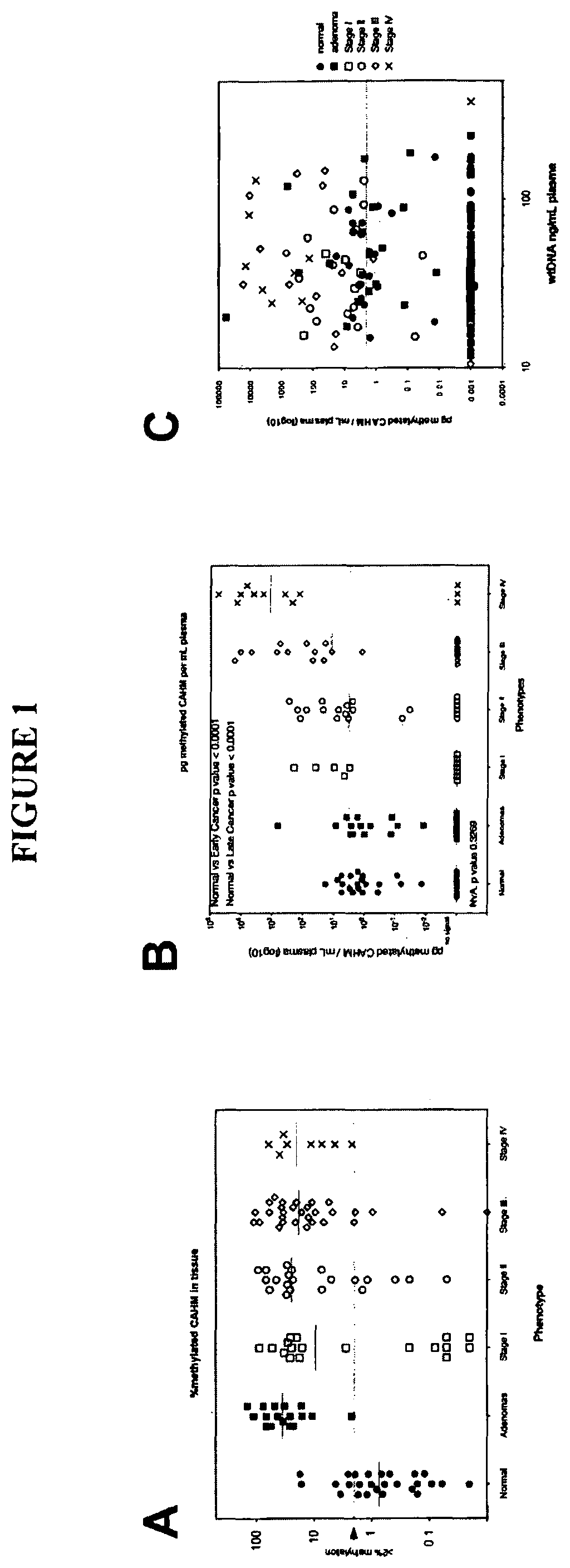
[0273]Tissue DNA samples were acquired through a commercial specimen bank (BioServe, @, US) and a tertiary referral hospital tissue bank in Adelaide, Australia. Blood plasma specimens were acquired from a commercial specimen bank (Proteogenex, Culver City, Calif.) and a tertiary referral hospital in Adelaide, Australia. Blood specimens were classified as normal, adenoma or cancer based on colonoscopy results verified (where appropriate) by histopathology. This also identified the stage of the cancer. Peripheral blood was drawn into K3EDTA VACUETTE blood tubes (Greiner-One, Monroe, N.C.) and transported to the processing laboratory on wet ice. Whole blood was centrifuged at 1,500 g (4° C.) for 10 minutes within 4 hours of blood draw and plasma was collected. The plasma was centrifuged for a second time at 1,500 g (4° C.) for 10 minutes, where after the plasma was collected and stored at −80° C. until further use.

Tissue DNA Extraction & Bisulfit...


PUM

PropertyMeasurementUnit
sizeaaaaaaaaaa
temperatureaaaaaaaaaa
volumeaaaaaaaaaa

Abstract

The present invention relates generally to a method of determining one or more probabilities of respective classifications of a neoplasm into one or more neoplastic categories. More particularly, the present invention relates to a method of determining the probability of classification of a large intestine neoplasm into one or more categories selected from adenoma, stage I, stage II, stage III or stage IV by screening for changes to the methylation levels of a panel of gene markers, including BCAT1, IKZF1, IRF4, GRASP and/or CAHM. The method of the present invention is useful in a range of applications including, but not limited to, those relating to the diagnosis and/or monitoring of colorectal neoplasms, such as colorectal adenocarcinosis.

Description

PRIOR RELATED APPLICATIONS[0001]This application is a U.S. national phase patent application under 35 U.S.C. 371 of International Patent Application No. PCT / AU2013 / 000519, filed May 17, 2013, which claims the benefit of priority to the U.S. Provisional Application No. 61 / 648,821, filed May 18, 2012, each of which is incorporated herein by reference in its entirety.FIELD OF THE INVENTION[0002]The present invention relates generally to a method of classifying a large intestine neoplasm and, in particular, classifying a colorectal neoplasm in a mammal. The present invention more specifically provides a method for assessing the probability that a large intestine neoplasm is a premalignant neoplasm, an early stage malignant neoplasm or a late stage malignant neoplasm. The method of the present invention is based on screening for modulation in the DNA methylation levels of one or more gene markers in blood samples from said mammal.BACKGROUND OF THE INVENTION[0003]Colorectal cancer include...

Claims


Application Information

Patent Timeline
Patent Type & Authority Patents(United States)
IPC IPC(8): C12Q1/6886G01N33/574
CPCC12Q1/6886G01N33/57446C12Q2523/125C12Q2600/112C12Q2600/154G01N2800/06G01N2800/50G16H50/30
Inventor LAPOINTE, LAWRENCE CHARLESPEDERSEN, SUSANNE K.BAKER, ROHANGAUR, SNIGDHATHOMAS, MELISSA
Owner CLINICAL GENOMICS PTY LTD
Why Eureka
Try Eureka