Medical apparatus and its visualisation

a medical equipment and visualisation technology, applied in the field of medical equipment and its visualisation, can solve the problems of difficult to locate the precise position of an introduced structure, difficult to read images, and inability to clearly visualise all of the important or relevant information in a single type of image, so as to improve the accuracy or sensitivity of the determined location, improve the visibility of the structure, and improve the accuracy of the determined location

Active Publication Date: 2021-09-21
UNIV COURT OF THE UNIV OF DUNDEE
View PDF43 Cites 0 Cited by
  • Summary
  • Abstract
  • Description
  • Claims
  • Application Information

AI Technical Summary

Benefits of technology

[0020]Selection, extraction and / or transformation may be conducted so as to reduce processing demands, and enable images to be generated more rapidly or with less processing power. Selection, extraction and / or transformation may be conducted so as to improve the accuracy or precision of the calculation of the calculated image, for example by reducing the amount of noise in the data from which the calculated image data is generated.
[0121]Reciprocation of the mass assemblies along and around the principal axis results in concomitant motion of the probe assembly along and around the principal axis, in use. It has been found that rotation of the probe member along and around the principal axis reduces accretion and also reduces the forces which must be applied in order to penetrate tissues of a subject. This is of particular importance for low amplitude reciprocation and in use of delicate probe members probe to bending.

Problems solved by technology

However it may not be possible to clearly visualise all of the important or relevant information in a single type of image.
For example, it can be difficult to locate the precise position of an introduced structure, such as a needle or a medical device in images optimised to show the features of soft tissues.
Displaying different types of images side by side or overlaid may make the images difficult to read, particularly when viewing changing real-time images or may obscure important information and there is a need for methods and apparatus to improve visualisation of information in medical images.

Method used


Image

  • Medical apparatus and its visualisation
  • Medical apparatus and its visualisation
  • Medical apparatus and its visualisation

Examples


Embodiment Construction

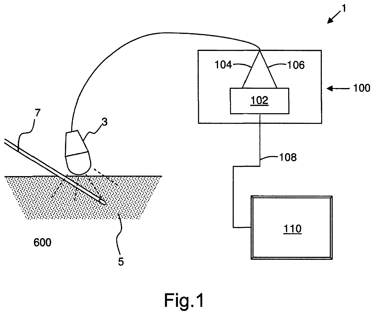
[0156]FIG. 1 shows a medical imaging system 1 which includes apparatus 100 for generating composite image data, according to an embodiment of the present invention. The system 1 has an imaging device 3 connected to a processing resource 102. A target region 5 of a subject, such as a patient, includes an introduced structure 7 and the imaging device is operable to acquire image data of the target region.

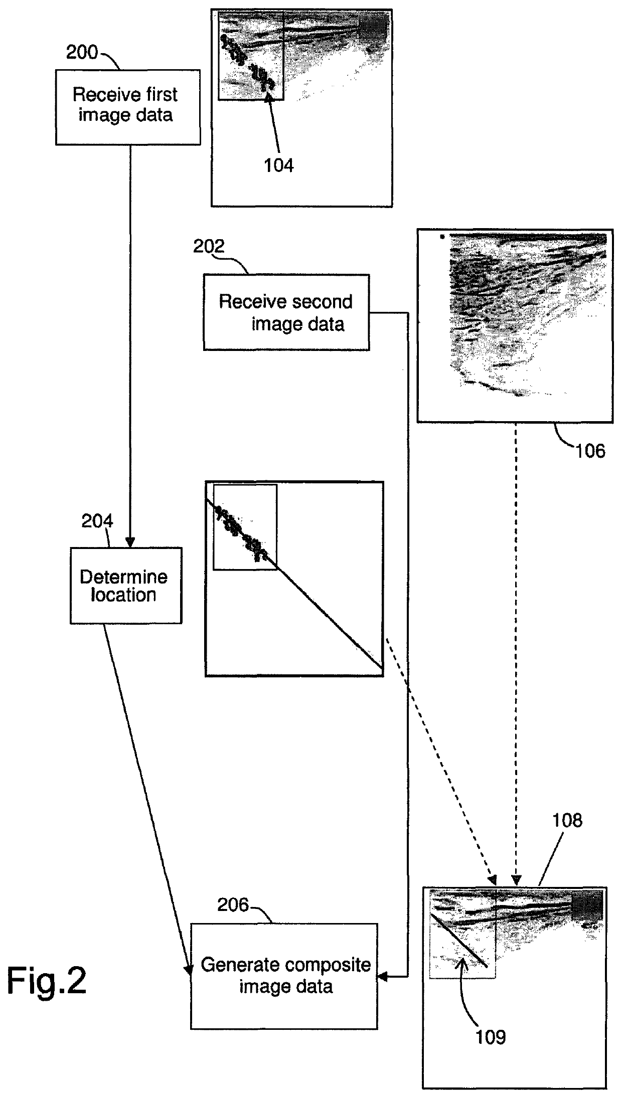
[0157]The processing resource is operable to receive first data 104 and second data 106, which comprises image data of the target region, from the imaging device. The processing resource is operable to generate composite image data 108, which is output to a display device 110, as described in further detail with reference to FIG. 2.

[0158]In the example shown, the imaging device is configured to acquire Doppler ultrasound image data (first image data) and B-mode ultrasound image data, and the imaging device 3 is an ultrasound probe and the introduced structure is an ultrasonically vibr...


PUM


Abstract

An ultrasonically actuated medical implement is used in improving medical interventions and in certain instances to the generation of image data, in particular data acquired during a medical intervention or procedure. The medical implement employs a piezoelectric element which causes reciprocation between first and second mass assemblies in order to ultrasonically actuate a probe member and improve the visibility of the probe member to imaging methods. The invention also concerns the visibility of structures in target regions to be imaged and how this may be enhanced using the medical implement of the invention.

Description

FIELD OF THE INVENTION[0001]The invention relates to improving medical interventions and in certain instances to the generation of image data, in particular data acquired during a medical intervention or procedure. The invention also concerns the visibility of structures in target regions to be imaged and how this may be enhanced.BACKGROUND TO THE INVENTION[0002]Medical images of a subject can include information which is difficult to interpret and the manner in which information is presented in medical images can be of critical importance.[0003]Different types of medical images are sensitive to different features in a region which is imaged. For example, MRI or ultrasound imaging is sensitive to soft tissues, X-ray imaging to denser bony tissues and Doppler imaging methods are sensitive to motion. However it may not be possible to clearly visualise all of the important or relevant information in a single type of image. For example, it can be difficult to locate the precise position...

Claims


Application Information

Patent Timeline
Patent Type & Authority Patents(United States)
IPC IPC(8): A61B17/34A61B17/32A61B8/08A61B8/00A61B10/02A61B90/00A61B17/00
CPCA61B17/3403A61B8/0841A61B8/463A61B8/488A61B8/5207A61B8/5246A61B17/320068A61B8/465A61B10/0233A61B2017/0011A61B2017/3413A61B2090/3929
Inventor SADIQ, MUHAMMADCOCHRAN, SANDYHUANG, ZHIHONGCORNER, GEORGEMCLEOD, GRAEMECARENA, PATRICK
Owner UNIV COURT OF THE UNIV OF DUNDEE
Why Eureka
Try Eureka