System and method for image-guided procedure analysis and training

a technology of image-guided procedures and training methods, applied in the field of system and method of image-guided procedure analysis and training, can solve the problems of increasing patient risk in these procedures, not decreasing, and unable to determine the exact position of the tip of the interventional instrumen

Active Publication Date: 2022-08-02
EZONO
View PDF15 Cites 0 Cited by
  • Summary
  • Abstract
  • Description
  • Claims
  • Application Information

AI Technical Summary

Benefits of technology

[0014]Thus with the invention, the storage and further processing of position data of the interventional instrument during the procedure allows consistent quantitative measures to be obtained relating to the quality of the procedure. For example, if the number of significant trajectory readjustments of the interventional instrument exceeds a small predefined threshold, for example two to four readjustments, this indicates that the clinician is hacking about in the patient, which can lead to pain, haematoma and infection. Being able to count the number of trajectory readjustments provides a reliable consistent measure which allows meaningful performance monitoring and meaningful comparisons between clinicians.
[0015]In an in-plane procedure where the interventional instrument is intended to advance in the imaging plane, determining the number of times the tip of the interventional instrument leaves the imaging plane gives a measure of the quality and skill level of the user, and determining the distance advanced by the tip of the instrument while it is out of the plane indicates whether the clinician was advancing the instrument while effectively blind to its position. This is dangerous as it can lead to complications, such as accidental puncturing of a blood vessel. Similarly, in an out-of-plane procedure where the interventional instrument is advanced at an angle to the imaging plane, counting the number of times the interventional instrument passes through the imaging plane, and the distance advanced, again provides a reliable measure of the skill level of the clinician and level of risk of the procedure.
[0021]Preferably the data store is configured to record the stored data with the identity of the operator of the procedure (clinician) and organisation, and preferably patient data. This allows statistical analysis of procedures based on identity of the clinician or identity of the organisation. This allows comparison between different user groups, monitoring changes in performance over time, benchmarking for levels of performance and the effectiveness of different training regimes.
[0022]The imaging system is preferably a freehand ultrasound imaging system, but the invention may also be used with an x-ray imaging system, magnetic resonance imaging system, PET imaging system as the recordal and processing of accurate position data of the interventional instrument allows the calculation of quantitative performance metrics regardless of the specific type of imaging system.
[0025]The system may further comprise an input for receiving a video image of the user conducting the image-guided interventional procedure. For example, a video camera such as a low cost webcam can monitor the user, providing useful additional information about their skill level. The system can include eye tracking of the user, thus providing more information about the user's skill level as it is important that the user focuses mainly on the image obtained by the imaging system rather than the interventional instrument itself.
[0026]The system may further comprise a physical phantom representing a patient's anatomy, this being useful for training purposes. Physical phantoms will have a known anatomy making it easy to perform image segmentation and provide more accurate quantitative measures of the trajectory and positioning of the interventional instrument relative to the anatomy. The physical phantom may be internally instrumented with a video camera (video endoscope) which provides additional video confirmation of whether the interventional instrument is in the correct part of the phantom's anatomy.

Problems solved by technology

In general, it is very difficult for the user to determine the exact position of the tip of the interventional instrument and thus to be sure whether it is in the desired place, for example adjacent a nerve, or whether it has undesirably penetrated something else, for example a blood vessel.
This has brought the benefits of ultrasound to many more patients through a wider base of non-traditional clinician users, but there has been a challenge in educating the non-traditional clinician users, and evaluating their skills.
However, excursions of the needle tip from the imaging plane mean that the tip becomes invisible in the ultrasound image, and advancing the needle in this state is potentially dangerous as the clinician does not know what is being penetrated.
If the needle tip crosses the plane it can be dangerous as it means the needle tip could puncture the back side of a vein which it is supposed to be in, could progress to puncture an artery which are typically close to veins or in a worst case could puncture the pleura and therefore cause pneumothorax.
If the skills of co-ordinating the movement of the interventional instrument and the imaging probe are not learnt correctly, patient risk can increase in these procedures, instead of decreasing.
The tools used for training are generally limited to presentation materials for theory, live model scanning for learning sono-anatomy, physical phantoms of all shapes, sizes and composition on which clinicians can learn needling techniques, and sometimes the use of cadavers for needling practice.
Despite these efforts, there is no clear evidence about the level of competence of users of image guided procedures.
Another problem is that it is expensive to devote point of care ultrasound apparatus to training.

Method used


Image

  • System and method for image-guided procedure analysis and training
  • System and method for image-guided procedure analysis and training
  • System and method for image-guided procedure analysis and training

Examples


Embodiment Construction

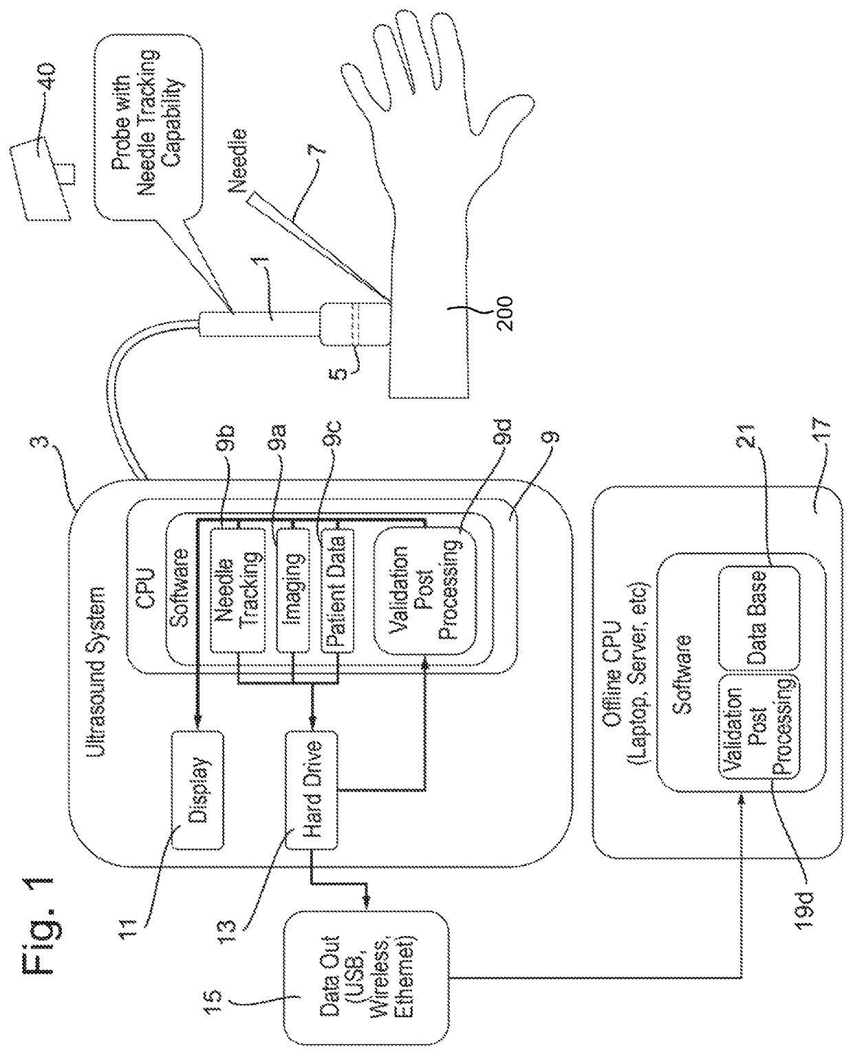
[0043]FIG. 1 schematically illustrates an embodiment of the invention applied to an ultrasound image-guided procedure using a system combining ultrasound imaging and magnetic tracking, such as disclosed by WO-A1-2013 / 034175 (eZono AG). As illustrated in FIG. 1, the imaging system comprises a freehand ultrasound imaging probe 1 which is controlled by and supplies ultrasound data to a combined ultrasound and position detection system 3. The ultrasound imaging probe 1 also includes a plurality of magnetic field detectors in a magnetic detector array 5 which detect the magnetic field from the interventional instrument 7 (in this case a magnetised needle), and supply magnetic field measurements to the combined ultrasound and position detection system 3.

[0044]The combined ultrasound and position detection system 3 includes a data processor 9, display 11 and data store 13 (which may be in the form of a disk drive or solid state hard drive). It optionally also includes a data output 15 (suc...


PUM


Abstract

A system and method for quantitative validation of the quality of image-guided interventional procedures combine image data on the procedure with position tracking of the interventional instrument and calculate quantitative measures such as excursions of the interventional instrument from a desired position or track, procedure times and measurements of distances advanced and withdrawn. The quantitative metrics may be graphically displayed and stored. Metrics obtained for multiple users may be stored and statistically processed to derive measures of performance applicable to individuals or groups of clinicians.

Description

CROSS-REFERENCES TO RELATED APPLICATIONS[0001]This application claims the benefit of European Patent Application No. 17176925.0, filed Jun. 20, 2017, the entire content of which is herein incorporated by reference.BACKGROUND[0002]The present invention relates to the field of image-guided procedure analysis, particularly to systems and methods for analysis and quality validation of image-guided interventional medical procedures and to apparatus for training clinicians in image-guided interventional medical procedures.[0003]There are numerous medical procedures that involve the insertion of an interventional tool or instrument, such as a needle, cannula, catheter, stylet, scalpel or other medical or surgical instrument, into a subject's body, e.g. minimally-invasive surgical procedures, regional anaesthesia, detection of bio-electrical signals, electrical stimulation for diagnosis or treatment, vascular access, fine needle aspiration, musculoskeletal injections and so on. In such proc...

Claims


Application Information

Patent Timeline
Patent Type & Authority Patents(United States)
IPC IPC(8): A61B5/055G09B23/28A61B6/12A61B6/03A61B5/06A61B8/08G06T7/10G06T7/20A61B17/34A61B34/20A61B8/00A61B34/10
CPCG09B23/286A61B5/055A61B5/062A61B6/037A61B6/12A61B8/0841A61B8/4254A61B8/463A61B17/3403A61B34/20G06T7/10G06T7/20G09B23/285A61B8/587A61B2017/3413A61B2034/107A61B2034/2055G06T2207/10016G06T2207/10088G06T2207/10104G06T2207/10116G06T2207/10132G06T2207/30004G06T2207/30241
Inventor DUNBAR, ALLANLEE, SUNGMO
Owner EZONO
Why Eureka
Try Eureka