Troponin I polypeptide fragments and uses thereof

a polypeptide and fragment technology, applied in the field of troponin i polypeptide fragments, can solve the problems of troponin i in vivo being unstable, unable to be used interchangeably amongcalibrators/controls, and undergoing degradation

Inactive Publication Date: 2002-09-12
SPECTRAL DIAGNOSTICS
View PDF0 Cites 13 Cited by
  • Summary
  • Abstract
  • Description
  • Claims
  • Application Information

AI Technical Summary

Problems solved by technology

Because of the differences in the methodology and components among the above-mentioned assays. and the calibrators / controls cannot be used interchangeably among assays.
This presents a major problem for the adoption of a universal standard and inter-laboratory comparisons of data.
Furthermore, troponin I in vivo is not a stable molecule and undergoes degradation by proteolytic enzymes present in the body.
Although it is somewhat protected from proteolysis by complex with troponin C, this complex is non-covalent and thus is subject to dissociation, rendering troponin I susceptible to proteolytic attack.

Method used


Image

  • Troponin I polypeptide fragments and uses thereof
  • Troponin I polypeptide fragments and uses thereof
  • Troponin I polypeptide fragments and uses thereof

Examples


example 1

Preparation of Recombinant Polypeptides Corresponding to Troponin I Fragments

[0042] The cardiac troponin I fragment 1-99 was cloned from the full-length human cardiac troponin I cDNA by polymerase chain reaction using specific primers designed to amplify the TnI 1-99 region. After confirmation by DNA sequencing, the cDNA was then inserted into a pET expression vector. E. coli strain BL21(DE3) was transformed with the resulting construct and the selected clones were grown in LB medium for protein production. Modifications of certain codons were made to facilitate expression, without altering the amino acid sequence. TnI 1-99 was isolated from E. coli cells using an affinity column coupled with TnI antibodies recognizing the 1-99 region.

[0043] The E. coli strain was deposited on Jul. 27, 1998, with the American Type Culture Collection. 10801 University Blvd., Manassas, Va. 20110-2209 and accorded Deposit No. 98824.

example 2

Epitope Mapping of Troponin I Fragments

[0044] The troponin I fragment described in Example I, as well as a recombinant, full-length troponin I molecule, additionally containing six N-terminal amino acids (as described in copending application Ser. No. 08 / 862,613 and Ser. No. 08 / 961858, both incorporated herein by reference), were assayed in the Stratus(R), Access(R), and Opus(R) assays, with the following qualitative results:

1 Stratus(R) Access(R) Opus(R) SEQ ID NO: 2 + - + (N-terminal 99 amino acids) Full-length troponin I plus an + + + additional 6 N-terminal amino acids

[0045] These data demonstrate the differential recognition of the troponin I epitopes by the various assay formats. Each of the assays employ two antibodies, a capture antibody, and a detector antibody. All three assay recognized full-length troponin I. The Stratus(R) and Access(R) assays employ two monoclonal antibodies. The latter recognizes the C-terminal portion of troponin I and thus was unable to detect the f...

example 3

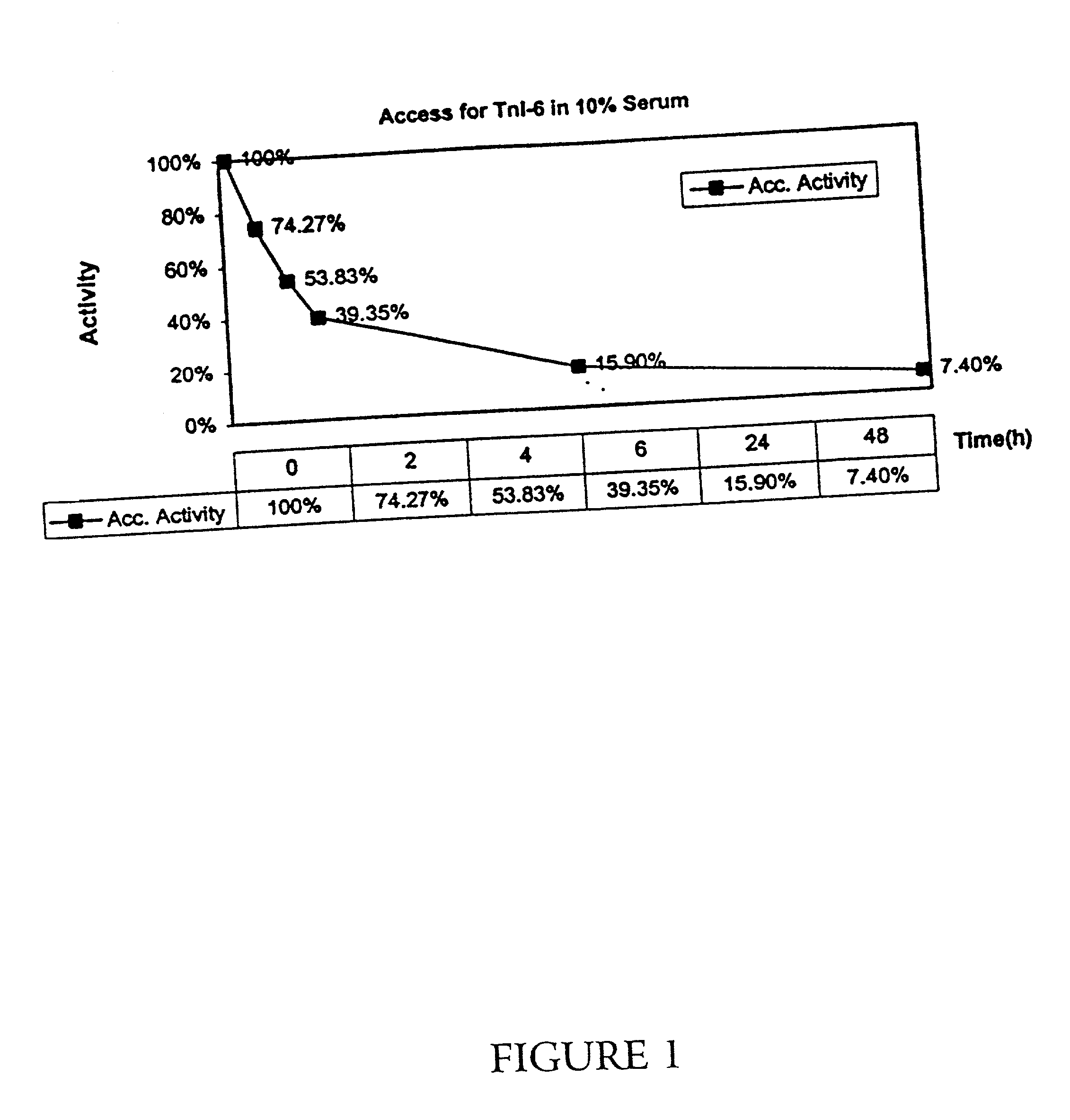
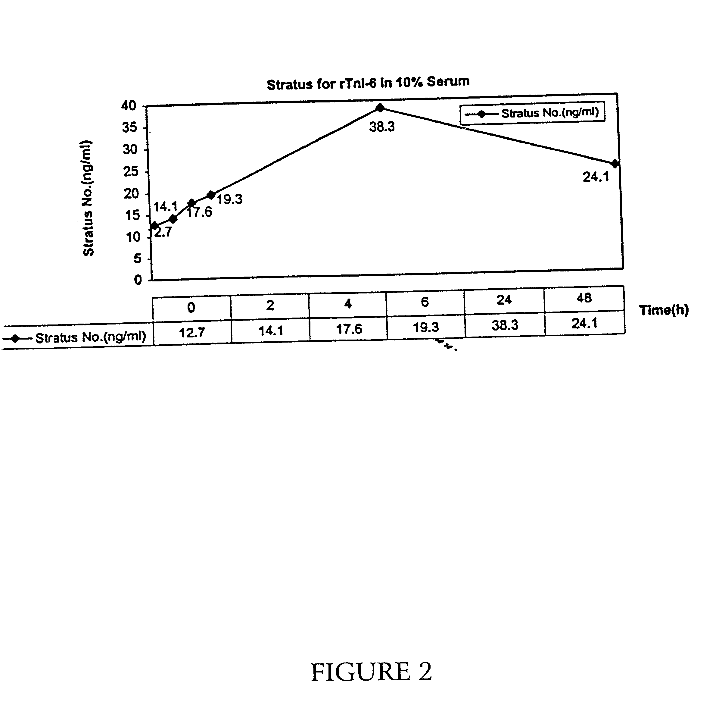
[0046] A recombinant, full-length troponin I molecule, additionally containing six N-terminal amino acids (as described above) was incubated in 10% human serum for 24 hours at room temperature. Samples were taken at 0, 2, 4, 6, 24, and 48 hours, and the level of troponin I was determined by immunoassay by the Access(R) and the Stratus(R) assavs for troponin I.

[0047] As shown in FIG. 1, the level of troponin I detected by the Access(R) assay decreased with time, with only 75% of the initial troponin I level present after 2 hours, and only 40% present after 6 hours. At 24 hours, only 16% was present. As described in above, the Access(R) recognizes the C-terminus of troponin I. As shown in FIG. 2. however, when the Stratus(R) assay (which detects a region near the N-terminus) is used on the same samples, an initial increase in detectable troponin I levels is apparent, with an apparent three-fold increase in levels at the 24-hour point, and the level subsequently falling. Thus, it appea...


PUM

PropertyMeasurementUnit
pHaaaaaaaaaa
stabilityaaaaaaaaaa
sizeaaaaaaaaaa

Abstract

Polypeptides corresponding to stable, circulating degradation products of troponin I (TnI) are described. The fragments comprise a sequence of the N-terminus of native cardiac TnI with 95-115 amino acid and additionally include fragments lacking about the 20-30 N-terminal amino acids. Utilities of these fragments and antibodies thereto include sensitive detection of myocardial infarction and purification of antibodies sensitive to the detection of troponin I degradation products.

Description

FIELD OF THE INVENTION[0001] This invention relates to new polypeptides corresponding to human cardiac troponin I N-terminal fragments and their uses as immunogens, affinity reagents, and standards for the preparation and calibration of improved immunoassays for troponin I.BACKGROUND OF THE INVENTION[0002] Early and accurate assessment of suspected acute myocardial infarction is critically dependent on the sensitive and specific detection and quantitation in blood, serum or plasma of released cardiac muscle intracellular components in order to distinguish a potentially lethal event in need of emergency measures from non-life threatening conditions such as angina and non-cardiac chest pain such as dyspepsia. Early electrocardiographic changes are neither adequately specific nor sensitive, and the medical profession has come to rely on serum biochemical markers of cardiac tissue injury for early diagnosis. Initially, the serum markers creatine kinase (CK) and specifically the cardiac ...

Claims


Application Information

Patent Timeline
Patent Type & Authority Applications(United States)
IPC IPC(8): A61K38/00C07K14/47C12N15/12
CPCA61K38/00C07K14/4716
Inventor SHI, QINWEILIU, SHIGUILING, MINGFU
Owner SPECTRAL DIAGNOSTICS
Why Eureka
Try Eureka