

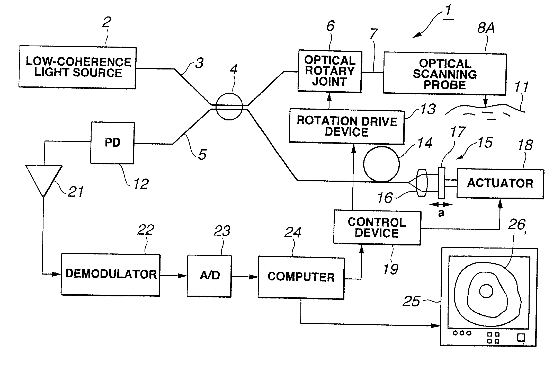
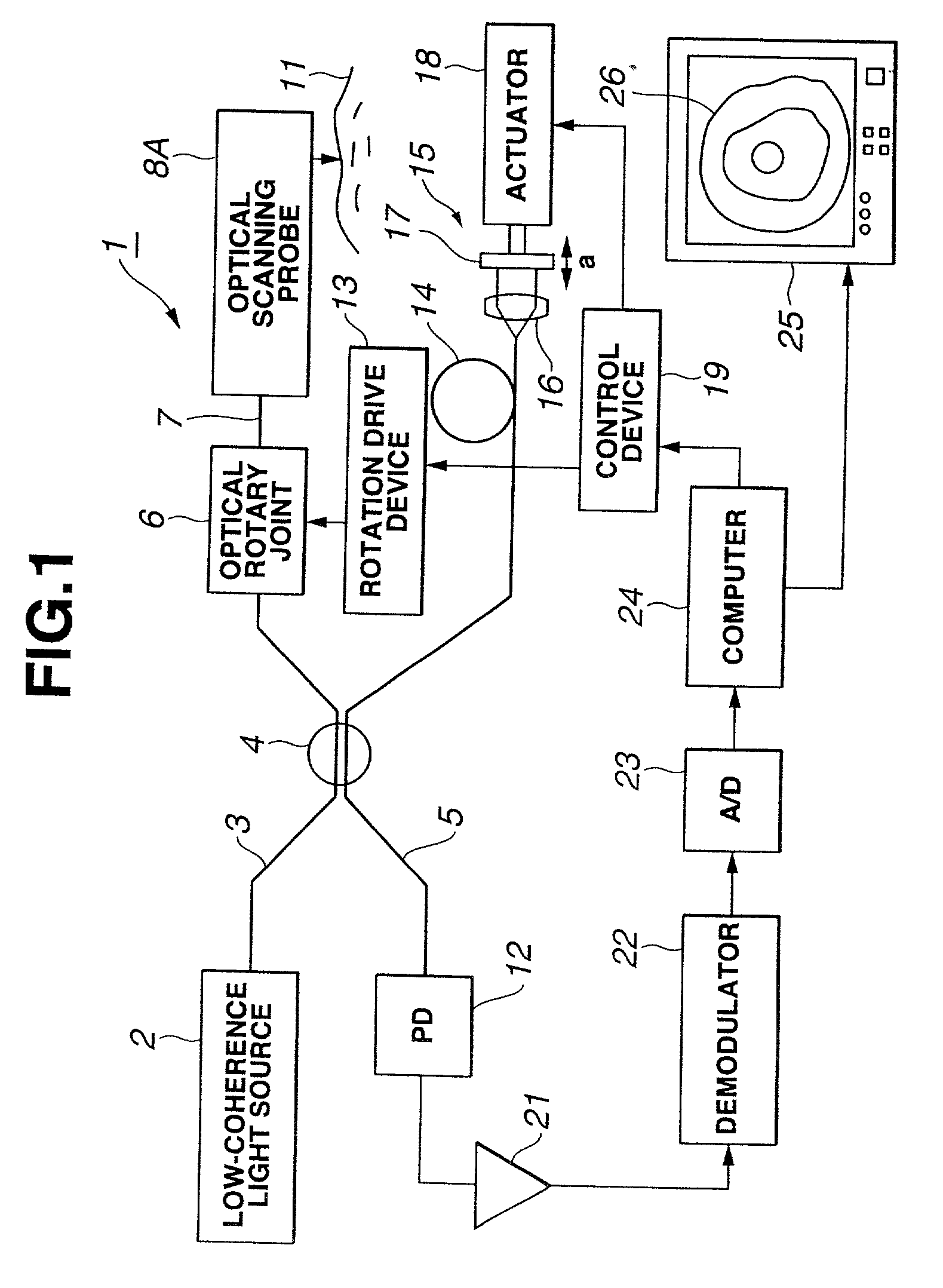
Optical scanning probe device using low coherence light
a low-coherence light and optical scanning technology, applied in the field of optical scanning probe devices, can solve the problems of difficult observation at a given distance from living organ tissue, high cost and large size of laser light sources,
- Summary
- Abstract
- Description
- Claims
- Application Information
AI Technical Summary
Benefits of technology
Problems solved by technology
Method used
Image

Examples
sixth embodiment
[0213] The sixth embodiment according to the present invention will be described with reference to FIG. 16A and FIG. 16B.
[0214] As shown in FIG. 16A and FIG. 16B, in an optical probe 8H according to the present embodiment, the tip opening of the sheath 42 is sealed watertight with the connection member 53 provided with the external thread member 53a on the outer perimeter of the tip, and a tip spacer 69 is screwed into the external thread portion 53a and, therefore, is attachable and detachable.
[0215] This tip spacer 69 is composed of an annular ring 69a, which has an outer diameter larger than the sheath 42 and which has a function of positioning, and a ring thread 69b, which is fixed to the inner perimeter of this ring 69a and in which internal thread portion is formed on the surface of the inner perimeter. Others are similar to those in the first embodiment.
[0216] Next, actions of the present embodiment will be described. When the tip spacer 69 is used as a positioning unit for k...
seventh embodiment
[0221] The seventh embodiment according to the present invention will be described with reference to FIG. 17 and FIG. 18.
[0222] As shown in FIG. 17, an optical probe 8I according to the present embodiment includes a probe main body 101 having the configuration similar to that of the prior example, and a balloon sheath portion 102 for storing this probe main body 101 in the inside.
[0223] In the probe main body 101, the flexible shaft 45, through which an optical fiber is inserted, is inserted through the transparent sheath 42, in which the tip is sealed watertight with a sheath seal member 77 and which has pliability, and the tip of this flexible shaft 45 is fixed to the housing 44 together with the tip of the optical fiber.
[0224] In this housing 44A, a GRIN lens is fitted facing to the tip of the optical fiber, the prism 52 is fitted at the tip face thereof and, therefore, the light can exit from and enter into the opening provided on the housing 44.
[0225] The rear end of the flexib...
eighth embodiment
[0237] The eighth embodiment according to the present invention will be described with reference to FIG. 19 to FIG. 21.
[0238] As shown in FIG. 19, an optical probe 8J according to the present embodiment is the optical probe 8I shown in FIG. 17 in which one end of an elastic tube 111 with the other end thereof being fixed to the tip of the outer sheath 104 is fixed on the sheath seal member 77 at the tip of the sheath 42.
[0239] This elastic tube 111 is in the shape of a tube having openings at the tip and the rear end, and is subjected to a shaping process beforehand in order that when no force is applied, an intermediate part of the tube body may protrude (evaginate) outside the radius and, therefore, a protrusion portion (evagination portion) 112 may be formed, as shown in FIG. 19 or FIG. 20.
[0240] The both ends of this elastic tube 111 are fixed watertight with string binding adhesion portions 113.
[0241] In the present embodiment, for example, the internal thread portion 105a of t...
PUM
| Property | Measurement | Unit |
|---|---|---|
| coherence length | aaaaa | aaaaa |
| coherence length | aaaaa | aaaaa |
| distance | aaaaa | aaaaa |
Abstract
Description
Claims
Application Information