Prostate visualization device and methods of use

a prostate and catheter technology, applied in the field of prostate cancer treatment, can solve the problems of long procedure, poor device mechanical and electrical performance, and poor accuracy of prostate cancer seeds,

Inactive Publication Date: 2005-01-06
NEOSEED TECH
View PDF49 Cites 11 Cited by
  • Summary
  • Abstract
  • Description
  • Claims
  • Application Information

AI Technical Summary

Benefits of technology

[0016] It is also an object of the present invention to provide methods and apparatus that provide reliable imaging of the bladder / prostate junction.
[0018] These and other objects of the present invention are accomplished by providing methods and apparatus for bladder / prostate junction imaging comprising a catheter having at least one distally deployable member that engages and defines the proximal region of the bladder. The deployable member is preferably constructed from a shape memory material that forms a petal-shaped configuration upon deployment. The deployable member is deployed within the patient's bladder, and may be retracted proximally to conform to the proximal wall of a patient's bladder. Gas pockets may be provided around the deployable member to enhance visibility with an ultrasound probe. Additionally, echo-contrast agent may be injected to the region to facilitate reliable imaging.
[0020] The deployment lumen may be used in combination with any of the drainage, contrast, and prostatic therapy lumens. In alternative embodiments, one or more catheter lumens may be configured to serve more than one function. For example, a single lumen catheter having only one distal outlet port may be provided whereby one or more proximal ports are in communication with the outlet port via the single lumen. In this embodiment, the single lumen of the catheter allows for passage of the deployable member, fluid injection, fluid drainage and delivery of prostatic therapy devices or therapeutic agents.

Problems solved by technology

The procedure is lengthy, especially if a lymph node dissection is simultaneously performed, and requires a hospital stay of 2-5 days.
Possible complications include impotence and urinary incontinence.
Although, when performed correctly, brachytherapy may provide several benefits when compared to prostate removal and other techniques, current apparatus and methods for delivering the seeds to target locations within the prostate are somewhat crude and are subject to practitioner error.
However, while Crowley may provide improved imaging, the device is mechanically and electrically complex, and is therefore costly.
The device described in that patent, however, does not improve on current junction imaging techniques.

Method used


Image

  • Prostate visualization device and methods of use
  • Prostate visualization device and methods of use
  • Prostate visualization device and methods of use

Examples


Embodiment Construction

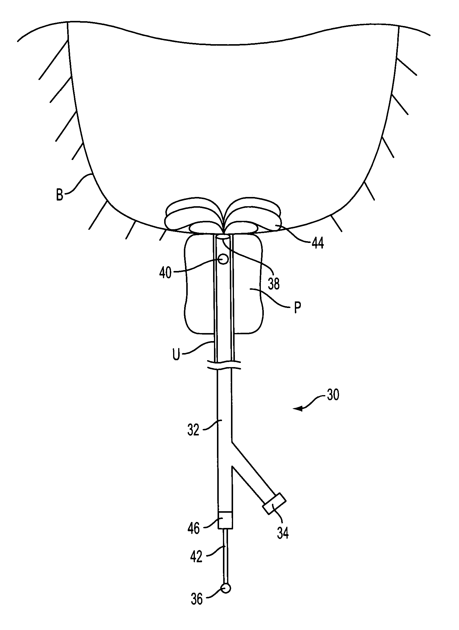
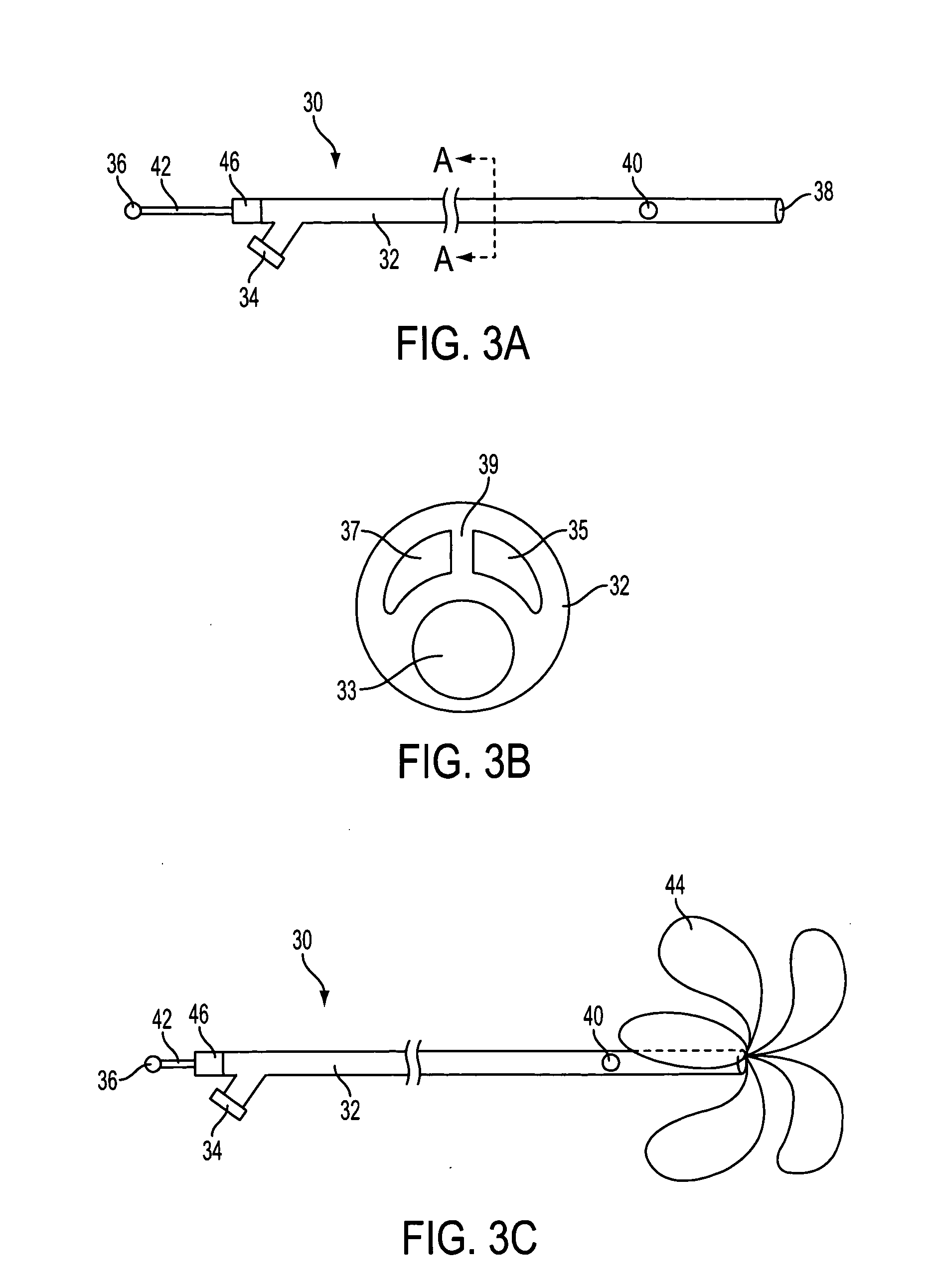
[0033] The present invention provides methods and apparatus for improved administration of brachytherapy. More particularly, the present invention provides a prostate visualization device comprising at least one distally deployable member that engages and defines the proximal wall of a patient's bladder. The device is preferably coupled to a catheter to facilitate imaging of the patient's bladder / prostate junction.

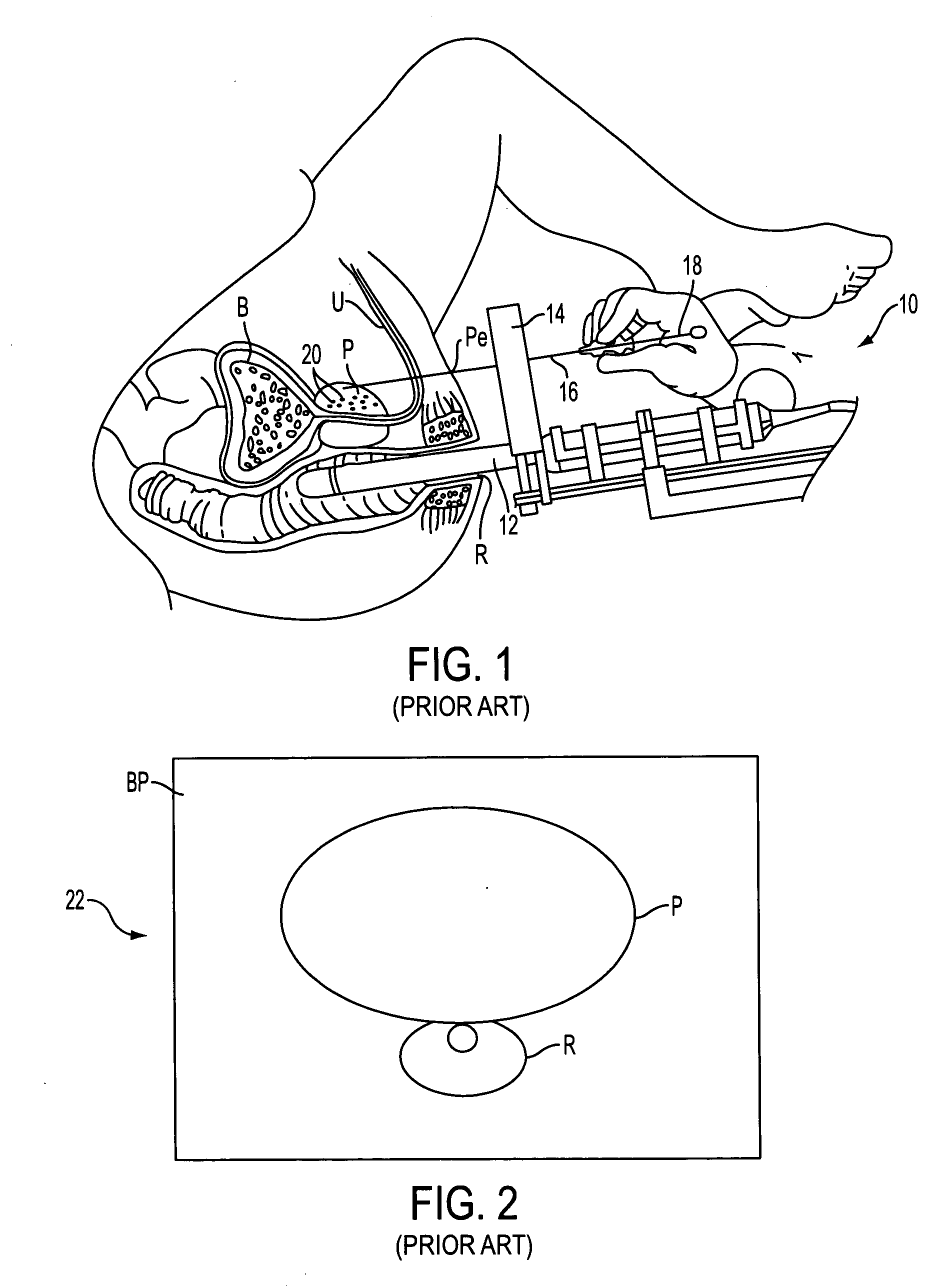
[0034] Referring to FIGS. 1 and 2, a prior art method of performing brachytherapy is described. The method and apparatus are as taught by Peter Grimm, DO, in a pamphlet entitled, “Ultrasound Guided Implantation of the Prostate: A Practical Review Course.” As seen in FIG. 1, brachytherapy apparatus 10 comprises transrectal ultrasound probe 12, guide block 14, needle 16, plunger 18, and radioactive seeds 20. Ultrasound probe 12 is advanced through a patient's rectum R to facilitate imaging of the patient's prostate P. Prostate P surrounds urethra U and is just proximal of b...


PUM


Abstract

Methods and apparatus are provided for improved administration of brachytherapy in the treatment of prostate disease. More particularly, a prostate visualization device is provided comprising a catheter coupled to at least one deployable member at the distal end of the catheter. The deployable member is preferably manufactured from a shape memory alloy having a petal-shaped configuration suitable for engaging and defining the proximal region of a patient's bladder. The deployable member may comprise tubing filled with air or other radiopaque agents to facilitate ultrasonic imaging of the deployable members near the bladder / prostate junction.

Description

REFERENCE TO RELATED APPLICATION [0001] The present application is a continuation-in-part of U.S. patent application Ser. No. 09 / 648,319, filed Aug. 25, 2000, and claims the benefit of the filing date of U.S. provisional application Ser. No. 60 / 295,408, filed May 31, 2001, the disclosure of which is incorporated herein by reference in its entirety.FIELD OF THE INVENTION [0002] This invention relates to improved apparatus and methods for the treatment of prostate cancer. More particularly, the present invention provides a prostate visualization device having at least one deployable member suitable for imaging the bladder / prostate junction, and methods of use. BACKGROUND OF THE INVENTION [0003] Excluding nonmelanoma skin cancers, prostate cancer is the most common cancer afflicting American men. The American Cancer Society estimates that over 180,00 new cases will be diagnosed in the U.S. in the year 2000 alone, and that nearly 32,000 people will die from the disease. Prostate cancer ...

Claims


Application Information

Patent Timeline
Patent Type & Authority Applications(United States)
IPC IPC(8): A61B8/00A61B8/08A61B8/12A61F2/958A61N5/10
CPCA61B8/0833A61B8/0841A61B8/12A61B8/481A61N2005/1011A61M25/1011A61M2210/1085A61N5/1001A61N5/1014A61M25/1002
Inventor HOGENDIJK, MICHAELGREEN, THOMAS C.GANDIONCO, ISIDRO M.BOUCHER, RYAN P.BELMAN, YURI
Owner NEOSEED TECH
Why Eureka
Try Eureka