Method and apparatus for visualization of 2D/3D fused image data for catheter angiography

a 3d reconstruction and fused image technology, applied in the field of 3d image postprocessing, can solve the problems of inability to verify the narrowing (stenose) caused by atherosclerotic deposits (plaque formation) on the inner wall, and require a relatively long post-processing time for 3d reconstruction, and achieve the effect of better orientation

Inactive Publication Date: 2005-01-20
SIEMENS AG
View PDF6 Cites 69 Cited by
  • Summary
  • Abstract
  • Description
  • Claims
  • Application Information

AI Technical Summary

Benefits of technology

[0018] An object of the present invention is to provide an image acquisition and processing technique, with which a 3D representation of blood vessels is enabled in the context of a contrast agent-free endovascular intervention, for better orientation in the navigation of an intravascularly inserted micro-catheter.

Problems solved by technology

The intravascular insertion of a micro-catheter for endovascular therapy of a blood vessel deformation, as well as the navigation of this catheter within the blood vessel system to be treated, is a difficult and demanding procedure, since the catheter must be guided through a number of vessel branchings.
Moreover, with this examination method vessel narrowings (stenoses) caused by atherosclerotic deposits (plaque formation) on the inner walls cannot be verified, or can be verified only indirectly.
Conventional reconstruction methods enable a 3D representation of larger vessels, but require a relatively long post-processing time for 3D reconstruction from x-ray images acquired from various projection directions.

Method used


Image

  • Method and apparatus for visualization of 2D/3D fused image data for catheter angiography
  • Method and apparatus for visualization of 2D/3D fused image data for catheter angiography
  • Method and apparatus for visualization of 2D/3D fused image data for catheter angiography

Examples


Embodiment Construction

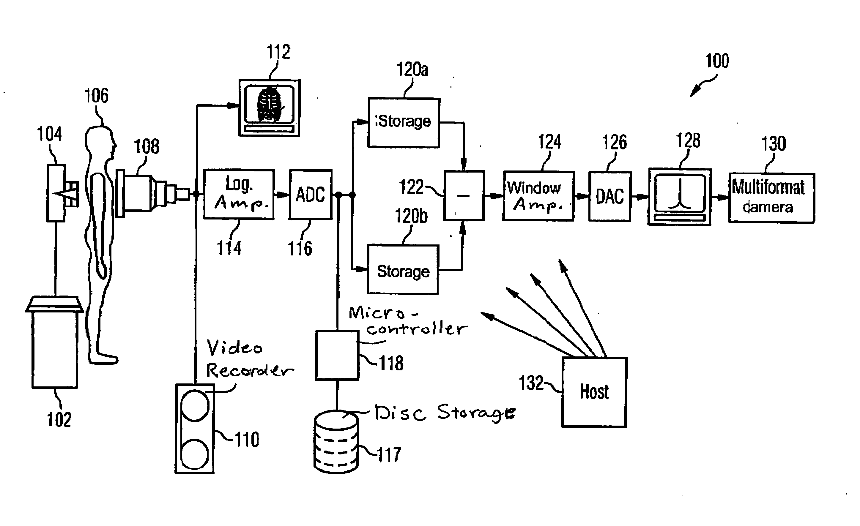
[0024] In the following, the invention imaging method as illustrated in FIG. 3 is first described in detail. The functioning of the system components depicted in FIG. 1 is subsequently explained.

[0025] As an exemplary embodiment of the invention, a computer-aided imaging and image post-processing method is described for angiographic representation of aneurysms, angiomas and other arterio-venal malformations (AVM) of parts of the blood vessel system of a patient. A micro-catheter 306 (which can be inserted intravascularly) serves for the injection of an x-ray contrast agent. Before the beginning of an endovascular intervention, a contrast agent-intensified angiography pre-examination (S1) for two-dimensional representation of the parts of the blood vessel system to be treated is implemented from at least two different projection directions, and a 3D reconstruction method (S2) is applied for the purpose of acquisition of a first image data set (DS1) in order to obtain a three-dimensi...


PUM


Abstract

In an image post-processing method and apparatus for 3D visualization of 2D/3D fused image data for use in catheter angiography in an endovascular interventional procedure, upon forward movement of a micro-catheter through blood vessel in the interventional procedure, x-ray images are acquired from different projection directions and are subjected to a pattern recognition algorithm for edge-based segmentation of the image regions filled by the micro-catheter, with all remaining image regions being masked out. The segmented projection exposures are prepared by a 3D reconstruction algorithm to obtain an image data set for (pseudo-) three-dimensional representation of the micro-catheter. This image data set are intraoperatively registered and fused with an image data set acquired from an angiographic pre-examination for three-dimensional visualization of the vessel topography. The reconstructed 3D representation of the catheter is mixed into the three-dimensionally prepared representation of endovascular blood vessel sections to be treated.

Description

BACKGROUND OF THE INVENTION [0001] 1. Field of the Invention [0002] The present invention concerns a 3D image post-processing method of the type wherein an x-ray contrast agent is administered via a micro-catheter (catheter angiography), as well as a medical-technical apparatus for three-dimensional representation of sections of the blood vessel system of a patient that, in particular in the field of coronary angiography, can be used to diagnose diseases of the coronary vessels, as well as in the field of neuro-angiography for (pseudo-) three-dimensional vascular imaging in the framework of an endovascular intervention. [0003] 2. Description of the Prior Art [0004] For preventative treatment of coronary and cerebral aneurysms, angiomas and other arterial-venous malformations (AVM), endovascular interventions are ever more frequently employed today as an alternative to coronary or neurosurgical procedures. In the preliminary stages of an endovascular intervention, a contrast agent-in...

Claims


Application Information

Patent Timeline
Patent Type & Authority Applications(United States)
IPC IPC(8): A61B6/00A61B6/12G06T7/00G06T11/00
CPCA61B6/12A61B6/466A61B6/481G06T2207/30048A61B6/5235G06T7/0028A61B6/504G06T7/33
Inventor MITSCHKE, MATTHIASRAHN, NORBERTRITTER, DIETERSCHEUERING, MICHAEL
Owner SIEMENS AG
Why Eureka
Try Eureka