Systemic gene delivery vehicles for the treatment of tumors
- Summary
- Abstract
- Description
- Claims
- Application Information
AI Technical Summary
Benefits of technology
Problems solved by technology
Method used
Image
Examples
example 1
CD-expressing NSCs Retain Tropism for Intracranial Glioma
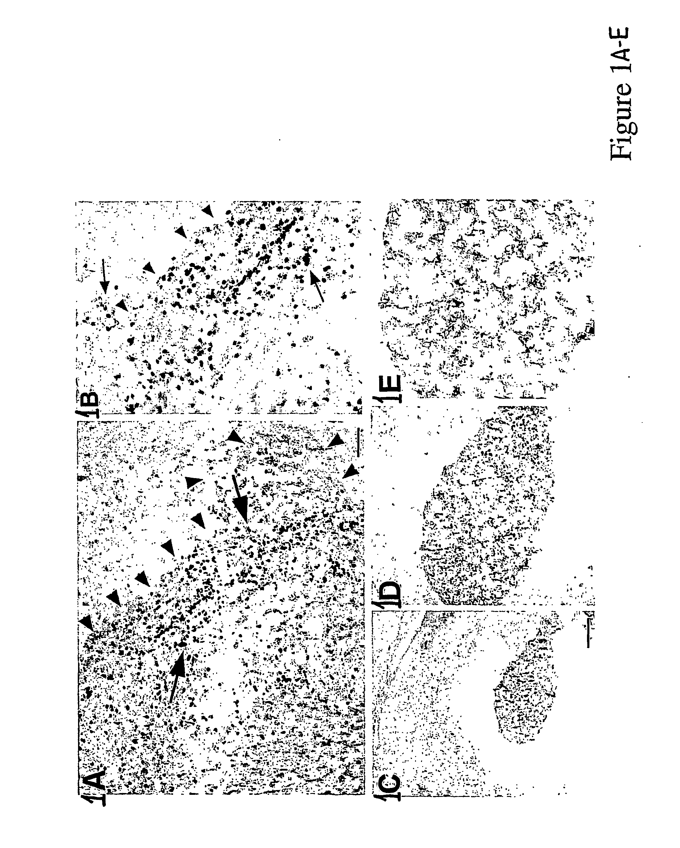
[0042] Several NSC lines, both human and murine, were transduced with a transgene encoding the bacterial pro-drug activating enzyme cytosine deaminase (CD) to determine if they retained their migratory, tumor-tracking properties. On day 0, adult CD-1 mice received stereotactically guided injections of CNS-1 cells (8×10⁴ in 2 μl PBS) into the right frontal lobe, as described above, and murine or human CD-NSCs (8×10⁴ in 2 μl PBS) into the left frontal lobe; Animals received daily injections of cyclosporine 10 μg / g) and were sacrificed on Day 7. The CD-transfected donor NSCs migrated across the corpus callosum and infiltrated the tumor in adult rodents.
[0043] As shown in FIGS. 1 A and 1 B, human CD-NSCs were found distributed throughout the tumor on the opposite hemisphere at day 7 after injection. These data support the premise that both murine and human NSCs modified to express a therapeutic bioactive agent (CD) behave in a s...
example 2
+CD-Expressing NSCs Produce Anti-Tumor Effect in Vitro
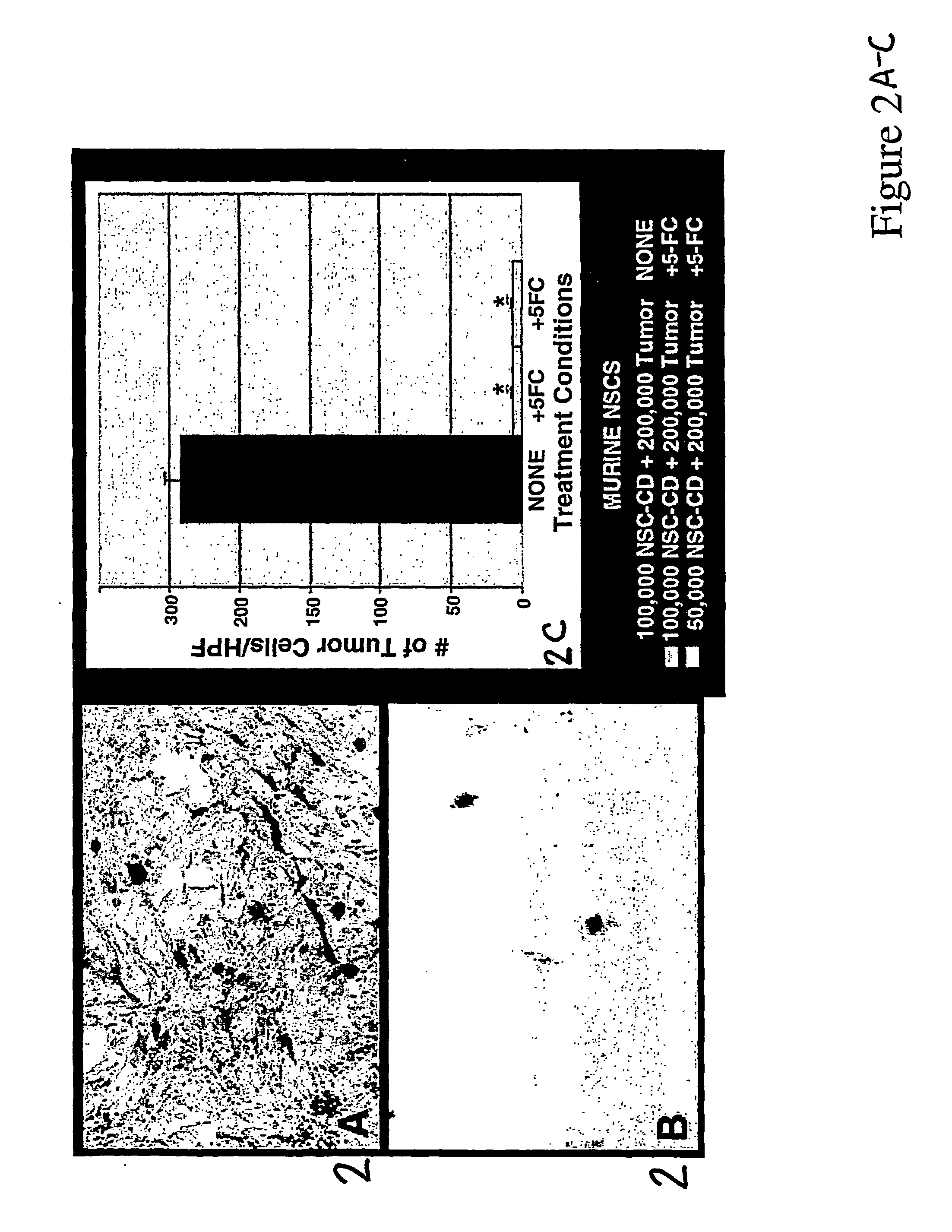
[0045] This experiment was performed to determine if modified CD-NSCs could effectively deliver therapy to tumor cells and produce a profound antitumor response. 200,000 CNS-1 glioma cells were plated into 10 cm Petri dishes. The following day (Day 1) media were replaced, and 50,000 or 100,000 CD-NSCs were added. On Day 2, media were replaced and 5-FC added (500 μg / ml). Control dishes included tumor-NSC co-cultures with no 5-FC treatment and tumor cell-only dishes with 5-FC treatment. Three days later, all plates were rinsed well with 1× PBS and fixed with 4% paraformaldehyde for 10 min at room temperature. Plates were stained with X-gal at 37° C. overnight to visualize NSCs and counterstained with neutral red to visualize tumor cells. Under high power, the numbers of tumor cells per field were counted. The tumor cell total was averaged from 20 random fields per plate. Error bars represent standard error of the mean.
[0046] Thes...
example 3
CD-Expressing NSCs Produce Anti-Tumor Effect in Vivo
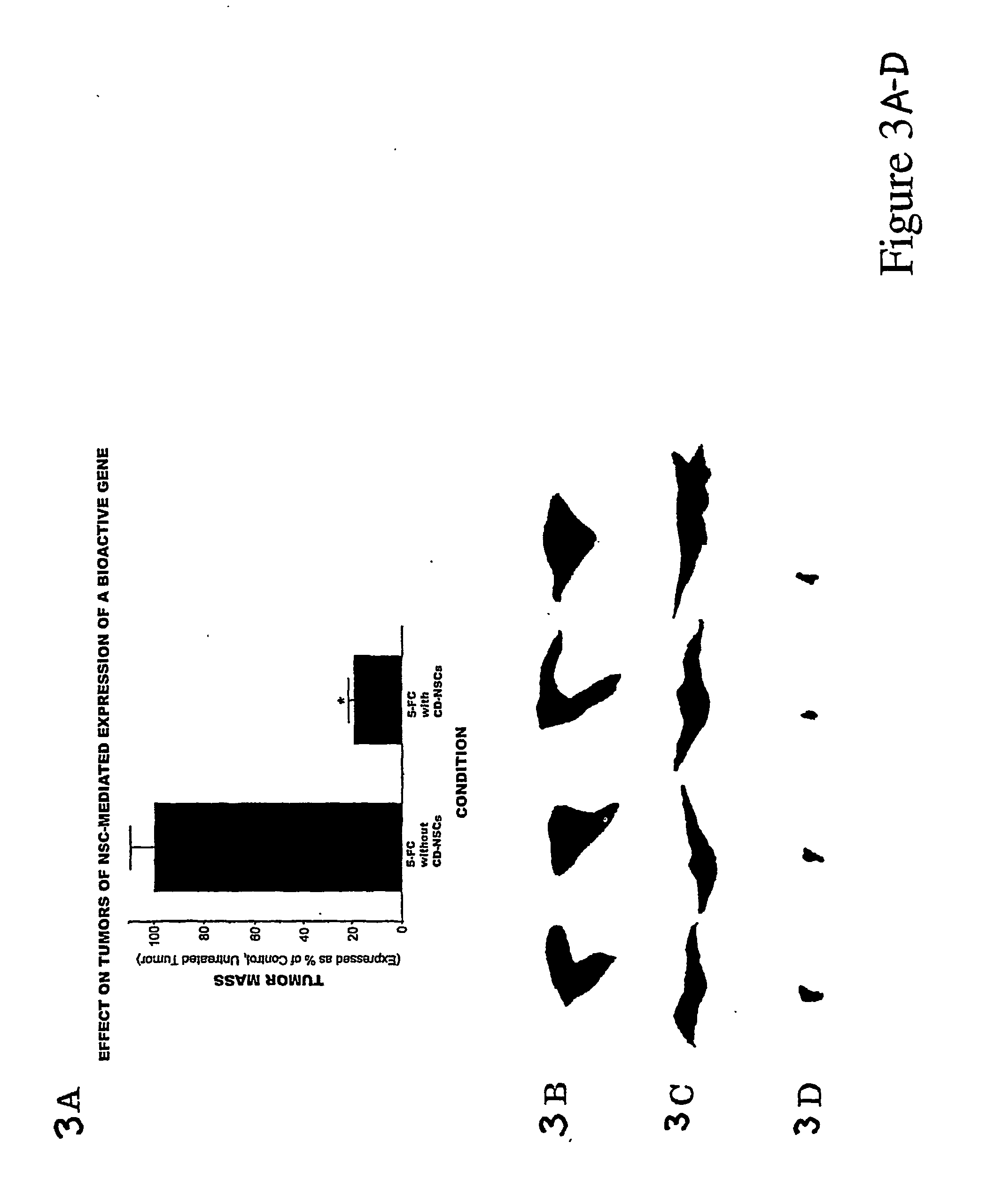
[0048] On Day 0, adult nude mice received stereotactically guided injections into the right frontal lobe of CNS-1 glioblastoma alone (7×10⁴ in 2 μl PBS), or CNS-1 cells mixed with CD-NSCs (7×10⁴ CNS-1 and 3.5×10⁴ NSCs in 2 μl PBS). Two days later treated animals received 10 intraperitoneal injections 900 mg / kg of 5-FC over a period of 10 days. Control animals received no 5-FC. Animals were sacrificed on day 13.
[0049] As shown in FIGS. 3A through 3D, animals receiving transplants of CD-NSCs and tumor cells followed by treatment with 5-FC, showed a significant reduction in tumor mass, as compared to the untreated animals.
PUM
| Property | Measurement | Unit |
|---|---|---|
| Therapeutic | aaaaa | aaaaa |
Abstract
Description
Claims
Application Information
 - R&D
- Intellectual Property
- Life Sciences
- Materials
- Tech Scout
- Unparalleled Data Quality
- Higher Quality Content
- 60% Fewer Hallucinations
Browse by: Latest US Patents, China's latest patents, Technical Efficacy Thesaurus, Application Domain, Technology Topic, Popular Technical Reports.
© 2025 PatSnap. All rights reserved.Legal|Privacy policy|Modern Slavery Act Transparency Statement|Sitemap|About US| Contact US: help@patsnap.com


