
Minimally invasive instrumentation for recovering tissue

a tissue and minimally invasive technology, applied in the field can solve the problems of insufficient specimen material for diagnosis, inability to allow a more advanced pathological investigation, and a risk of false positive, so as to improve the performance of tissue retrieval capture component cable assemblies, improve the tensional strength, and improve the effect of strength

Active Publication Date: 2005-02-10
COVIDIEN AG
View PDF1 Cites 75 Cited by
  • Summary
  • Abstract
  • Description
  • Claims
  • Application Information

AI Technical Summary

Benefits of technology

The present invention is addressed to apparatus for electrosurgically cutting about a tissue volume. Looking to minimally invasive devices incorporating tissue retrieval features, cable manipulating eyelet structures are employed at the tips of multiple leaf assemblies which are capable of withstanding bending stresses imposed during cutting movement through dense tissue. Formed integrally with these leaf assemblies, and retaining their thickness, the eyelet structures are twisted or bent into perpendicularity with respect to the faces of an associated leaf. While exhibiting increased but constant widthwise dimensions with minimized lengths, the structures remain desirably small in size but exhibits substantially improved structure integrity when subjected to surgical activity within the environment of very dense tissue.
As another object and feature of the invention, the performance of tissue retrieving capture component cable assemblies is substantially improved in terms of tensile strength at the very high temperature environment associated with a cable supported electrosurgical cutting arc. Such performance not only requires improved tensional strength at high temperature, but also requires the maintenance of proper mechanical deployment characteristics. This latter requirement essentially precludes substantial increases in nominal cable diameter. The improved strength at higher temperatures is achieved by employing cable with strands of only about a 1.4 mil diameter formed, for instance, of a type 316 stainless steel. Such improved strength is at an unexpected level. Published tensile strength at high temperature data for the type 316 material indicates only about a 30% strength improvement over type 304 stainless steel which was earlier employed to form the cable strands. However, the cable formed with type 316 stainless steel has been determined to be more than 104% stronger (a factor of 2.04) than the earlier multi-strand cable formed with type 304 material.

Problems solved by technology

With fine needle aspiration, there remains a risk of false-negative results, which most often occur in cases involving extremely fibrotic tumor.
In addition, after the procedure has been performed there may be insufficient specimen material for diagnosis.
Rather fragmented portions of tissue are withdrawn which do not allow a more advanced pathological investigation.
This limitation also is observed with respect to large core needle biopsies.
However, they still do not provide optimum pathological information.
In addition, while less expensive than open surgical biopsy, the ABBI has proven expensive compared to other biopsy techniques, and it has been noted that the patient selection for ABBI is limited by the size and location of the tumor, as well as by the presence of very dense parenchyma around the tumor.
Unacceptable increases in cable diametric size also would increase the power required for electrosurgical cutting.
Test experience with the capture components utilizing compressed porcine tissue has determined that, where the retrieval procedures encounter very dense breast tissue, the eyelets may fail by folding back.

Method used


Image

  • Minimally invasive instrumentation for recovering tissue
  • Minimally invasive instrumentation for recovering tissue
  • Minimally invasive instrumentation for recovering tissue

Examples


Embodiment Construction

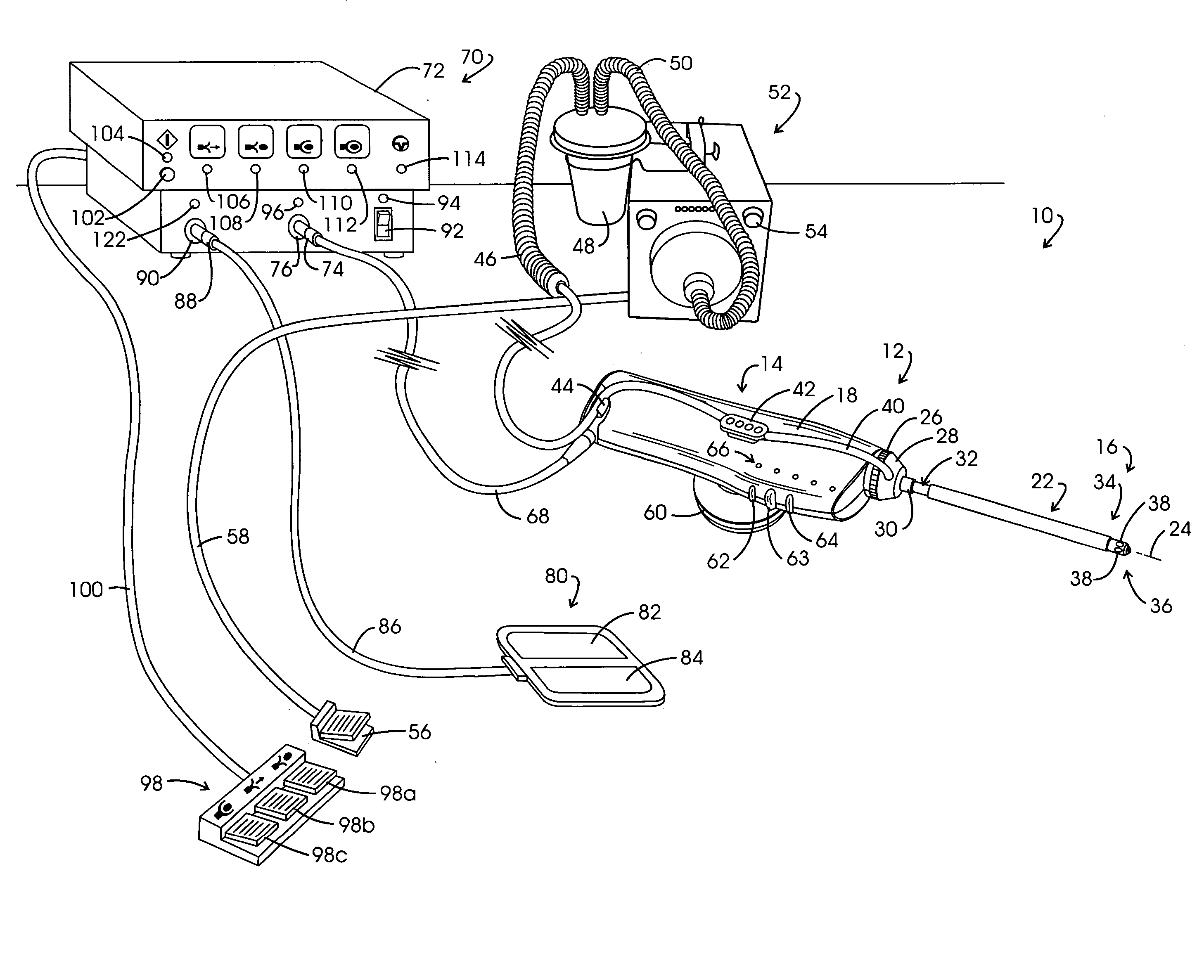
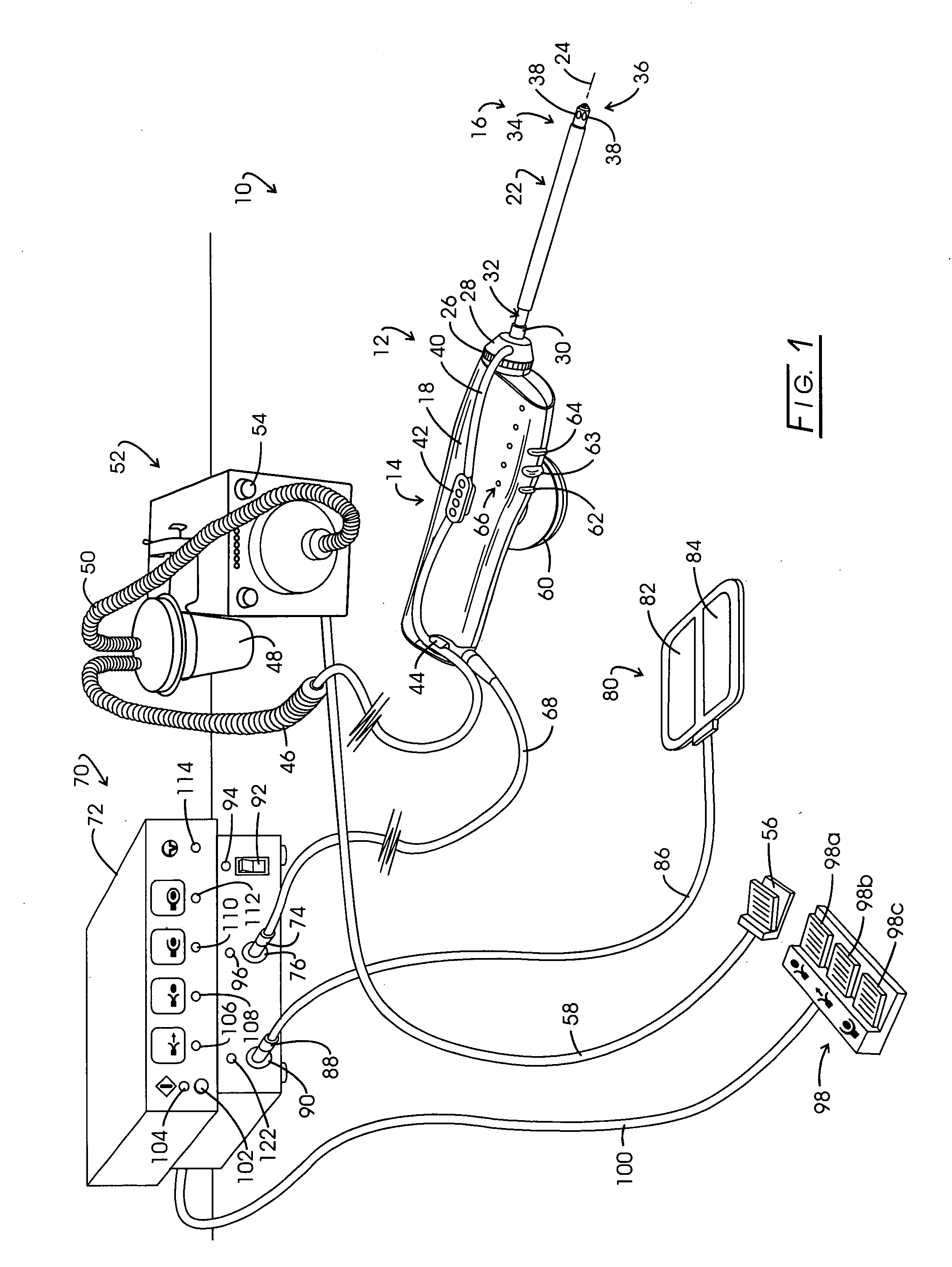
In the discourse to follow, computational data as well as test data taken with breast phantom materials are set forth. These data materials were developed in conjunction with investigations carried out with the noted tissue retrieval system marketed under the trade designation EN-BLOC®. Accordingly, that system is initially described.

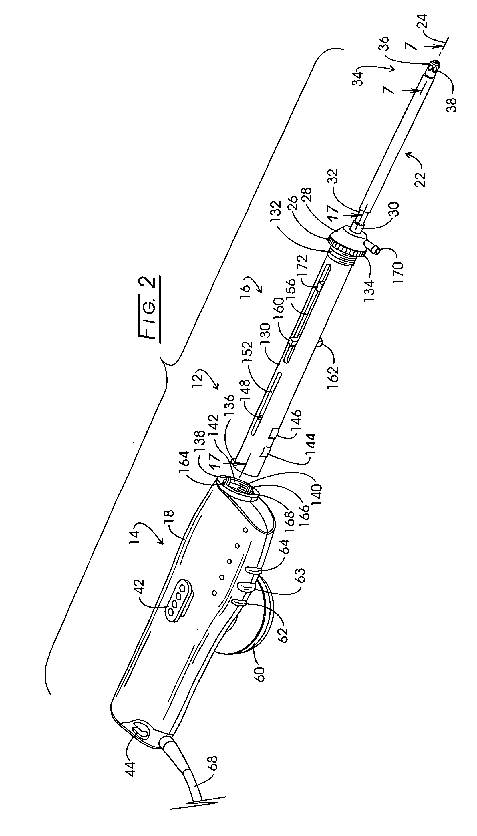
Referring to FIG. 1, the noted system for isolating and retrieving a target tissue volume or biopsy sample is illustrated in general at 10. System 10 comprises a tissue retrieval instrument represented generally at 12 which includes a reusable component represented generally at 14, sometimes referred to as a “handle”. Instrument 12 additionally includes a disposable component represented generally at 16, the rearward portion of which is removably mounted within the polymeric housing 18 of reusable component 14. The disposable component 16 is sometimes referred to as a “probe”.

Disposable component 16 includes an elongate cannula assembly or support ...


PUM


Abstract

The capture component of tissue retrieval apparatus is strengthened to improve its structural integrity when utilized within very dense tissue. Eyelet structures carrying pursing cable are improved through the utilization of slightly expanded constant widths with diminished lengths to avoid fold back phenomena. The pursing cables employed with the capture component exhibit more than a 100% improvement in tensile strength at high electrosurgical cutting temperatures through the utilization of strands having about a 1.4 mil diameter formed with a type 316 stainless steel.

Description

CROSS-REFERENCE TO RELATED APPLICATIONS STATEMENT REGARDING FEDERALLY SPONSORED RESEARCH Not applicable. BACKGROUND OF THE INVENTION The detection of tumorous lesions in the breast has progressed from early observation and palpation procedures to a variety of somewhat sophisticated imaging systems. A consequence of these advances in tumor detection is the identification of suspect tumor at an early stage in its development. Generally, at such early stages the suspect tumor may be somewhat small. Rather than resort immediately to an open surgical resection upon such early detection, practitioners generally carry out a preliminary, minimally invasive biopsy procedure. Such preliminary biopsy approaches are of importance, inasmuch as statistically, only 20% of these small tumors will be found to be malignant. Tumors determined to be benign have been left in situ with no excision. Over one million of these biopsies are performed in the United States each year, the procedure providing ...

Claims


Application Information

Patent Timeline
Patent Type & Authority Applications(United States)
IPC IPC(8): A61BA61B10/02A61B18/14A61B18/18
CPCA61B10/02A61B2018/144A61B2018/1407A61B18/148
Inventor EGGERS, PHILIP E.EGGERS, ANDREW R.EGGERS, ERIC A.JACOBS, DAVID
Owner COVIDIEN AG
Learn More