3D image processing apparatus

a technology of image processing and 3d, applied in the field of 3d image processing apparatus, can solve the problems of low utility of 3d-dsa images for surgical operation support, difficult to separate blood vessels from bones and soft tissues in a 3d-dsa image clearly

Active Publication Date: 2005-03-03
TOSHIBA MEDICAL SYST CORP
View PDF12 Cites 34 Cited by
  • Summary
  • Abstract
  • Description
  • Claims
  • Application Information

AI Technical Summary

Benefits of technology

[0005] It is an object of the present invention to separate blood vessels from bones clearly, and to display blood vessels in high definition.

Problems solved by technology

For this reason, the 3D-DSA images are low in utility for surgical operation support.
However, it is difficult to separate blood vessels from bones and soft tissues in a 3D-DSA image clearly.

Method used


Image

  • 3D image processing apparatus
  • 3D image processing apparatus
  • 3D image processing apparatus

Examples


Embodiment Construction

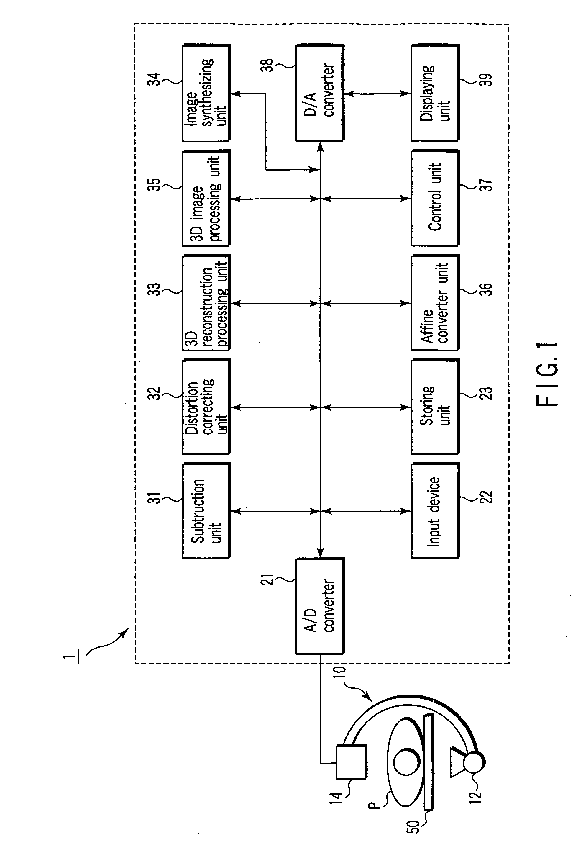
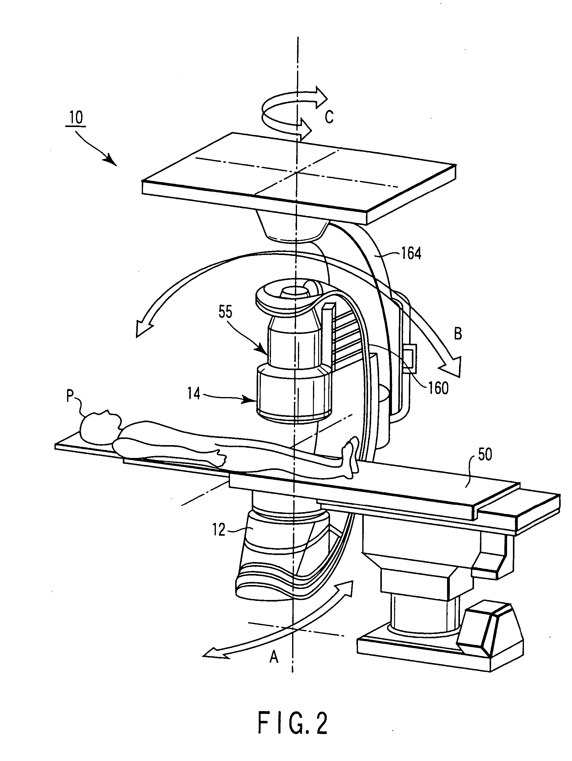
[0021] As shown in FIG. 1, a 3D X-ray diagnosing apparatus has an X-ray imaging mechanism 10 and 3D image processing apparatus 1. As shown in FIG. 2, the X-ray imaging mechanism 10 has an X-ray tube 12 and detection system 14. The detection system 14 is constituted by an image intensifier and TV camera. The detection system 14 may be formed from a flat panel detector. The X-ray tube 12 is mounted on a C-arm 160 together with the detection system 14. An object P to be examined on a top 50 of a bed is placed between the X-ray tube 12 and the detection system 14. The C-arm 160 is supported by a support 164 suspended from the ceiling. The C-arm 160 can rotate along three orthogonal axes A, B, and C.

[0022] The 3D image processing apparatus 1 includes a control unit 37 serving as a main component, an A / D converter 21, an input device 22, a storing unit 23, a subtracting unit 31, a distortion correcting unit 32, an affine converter 36, a 3D reconstruction processing unit 33, a 3D image pr...


PUM


Abstract

A 3D image processing apparatus comprises a storing unit storing mask images corresponding to projection directions associated with a subject contrast images corresponding to the projection directions, a subtracting unit generating subtraction images by subtracting the mask images from the contrast images, a reconstruction unit reconstructing first volume data from the mask images and reconstructs second volume data from the subtraction images, an image processing unit generating a first 3D image representing a bone structure and/or a soft tissue structure from the first volume data, and generates a second 3D image representing a contrasted blood vessel from the second volume data, an image synthesizing unit generating a synthetic image by synthesizing the first 3D image with the second 3D image, and a displaying unit displaying the synthetic image.

Description

BACKGROUND OF THE INVENTION [0001] 1. Field of the Invention [0002] The present invention relates to a 3D image processing apparatus. [0003] 2. Description of the Related Art [0004] A highly precise blood vessel structure can be extracted from a 3D-DSA image reconstructed from a plurality of DSA (Digital Subtraction Angiography) images corresponding to a plurality of projection directions. 3D-DSA images therefore are very useful for IVR support. In subtraction processing, however, the data of bones and soft tissues, other than contrasted blood vessels, are removed. 3D-DSA images therefore include no bone structure. For this reason, the 3D-DSA images are low in utility for surgical operation support. A 3D-DA image reconstructed from a plurality of DA (Digital Angiography) images corresponding to a plurality of projection directions includes a bone structure and blood vessel structure. An observer can therefore grasp the position of the blood vessel relative to the bone from the 3D-DA...

Claims


Application Information

Patent Timeline
Patent Type & Authority Applications(United States)
IPC IPC(8): A61B6/00A61B6/03G06T1/00G06T3/00G06T5/00G06T11/00G06T15/08H03F1/26H04N5/325
CPCA61B6/481A61B6/504G06T11/005G06T2211/404A61B6/584
Inventor OHISHI, SATORU
Owner TOSHIBA MEDICAL SYST CORP
Why Eureka
Try Eureka