Shielded dome resonator for mr scanning of a cerebrum

a dome resonator and cerebrum technology, applied in the field of mr scanning and imaging systems of cerebrum, can solve the problems of inefficient bird cages and tem coils, inability to provide power dissipation, and inability to provide patient comfort and imaging efficiency, so as to prevent parasitic coupling, improve sensitivity and electrical isolation, and improve imaging efficiency

Active Publication Date: 2005-03-17
GE MEDICAL SYST GLOBAL TECH CO LLC
View PDF8 Cites 17 Cited by
  • Summary
  • Abstract
  • Description
  • Claims
  • Application Information

AI Technical Summary

Benefits of technology

[0016] One of several advantages of the present invention is that it provides a dome resonator that is capable of performing high field magnetic resonance imaging and spectroscopy studies of a cerebrum with improved sensitivity and electrical isolation.
[0017] Another pair of advantages of the present invention is that in being dome shaped provides both patient comfort and imaging efficiency.
[0018] Furthermore, the present invention provides a dome resonator that is shielded, which prevents parasitic coupling to the surroundings, and therefore consequently prevents detuning of the resonator circuit and extraneous electrical losses.
[0019] Moreover, the present invention provides a dome resonator configuration such that distance between the shield and the resonator circuit decreases towards an apex of the dome resonator. In so doing, the present invention increases attenuation of field of the resonator near the apex where field strength is typically highest, thus providing a uniform field within the dome resonator. In providing a uniform field image quality is increased.

Problems solved by technology

Standard geometry of a coil of the shielded birdcage resonator type and of the TEM type provide a uniform RF B field, but can be claustrophobic in nature and are inefficient for imaging of just a cerebrum, since they bound a relatively large volume as compared to coils of the dome resonator type, which contain comparatively less tissue.
The birdcage and the TEM coils are inefficient due to their large size, which can cover a larger portion of a patient than is usually of interest.
Also, the birdcage and the TEM coils experience greater than necessary power dissipation and usually acquire data from regions of a body that are not necessarily of interest.
Coils of the dome resonator type are streamlined, patient friendly, and efficient, but produce a non-uniform RF B field.
The non-uniform B field increases in strength closer to the apex, due to bunching or close proximity of the conductive runners and concomitant bunching of magnetic flux lines.
The increase in field strength near the apex causes the field to be non-uniform and thus degrades image quality.
In addition to the above stated and associated disadvantages of the existing RF coil devices, the RF resonators are, as noted above, more and more likely to be subject to parasitic electromagnetic couplings to surrounding metallic objects as well as to radiation losses.
It is desirable to operate within increased static field strengths, such as 3.0 T and 4.0 T, instead of the conventional 1.5 T. Unfortunately, due to electrical properties of a patient, artifacts are increased at the higher static field strengths.
The result is the production of artifacts in the form of distorted image intensities, which are typically brightened at the centers of the images.

Method used


Image

  • Shielded dome resonator for mr scanning of a cerebrum
  • Shielded dome resonator for mr scanning of a cerebrum
  • Shielded dome resonator for mr scanning of a cerebrum

Examples


Embodiment Construction

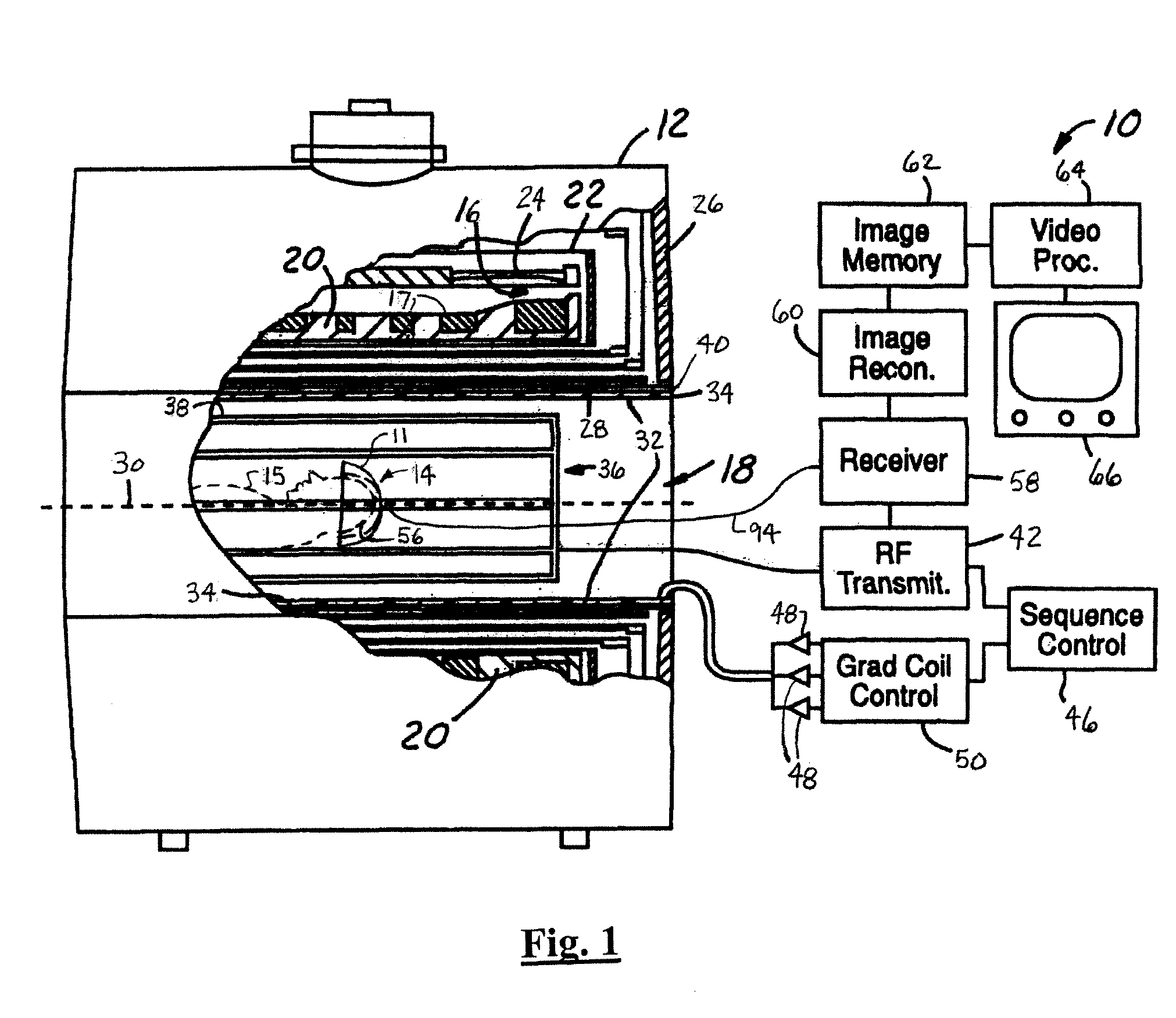
[0034] In the following figures the same reference numerals will be used to refer to the same components. While the present invention is described with respect to an apparatus and system for magnetic resonance (MR) and spectroscopy scanning and imaging of a cerebrum, the following apparatus and system is capable of being adapted for various purposes and is not limited to scanning or imaging of a cerebrum or to the following applications: magnetic resonance imaging (MRI) systems, magnetic resonance spectroscopy systems, and other similar applications known in the art.

[0035] In the following description, various operating parameters and components are described for one constructed embodiment. These specific parameters and components are included as examples and are not meant to be limiting.

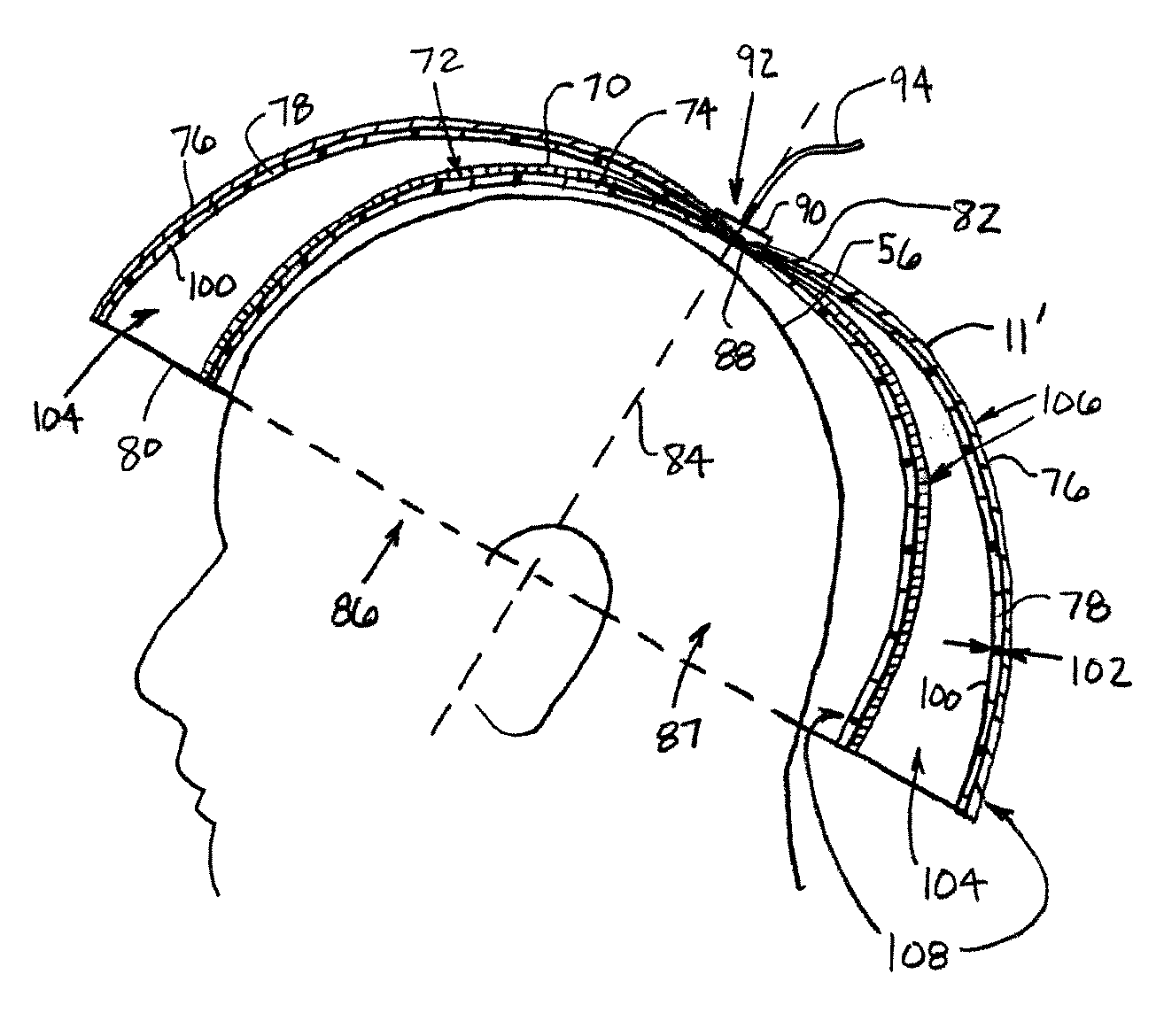
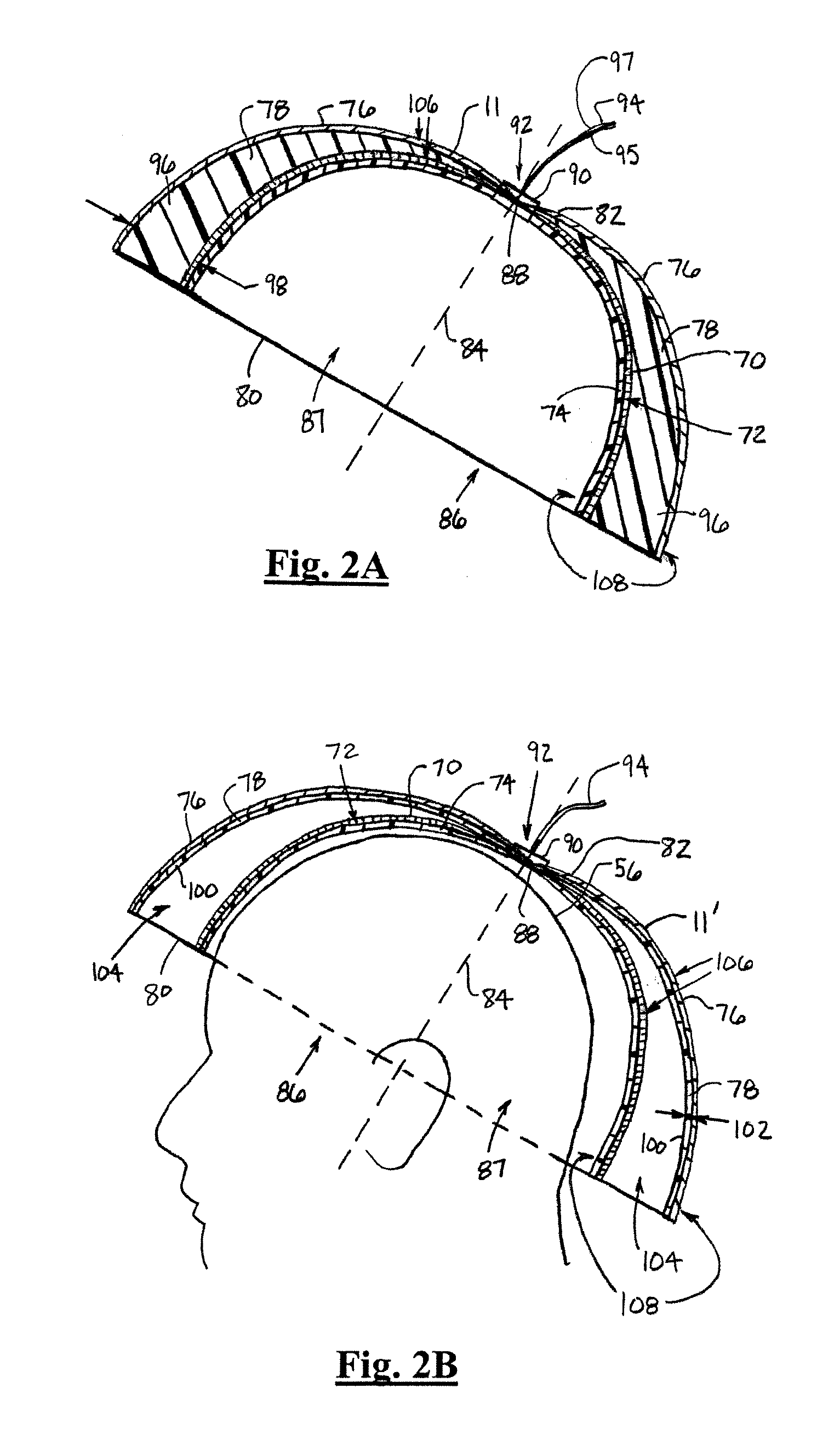
[0036] Also, in the following description the term “dome” refers to approximate shape of a device or object. The device or object has a center main portion with a first end and a second end. The c...


PUM


Abstract

A dome resonator (11) includes a resonator circuit (70) that excites and/or receives radio frequency magnetic resonance signals that emanate from a region of interest (14). The resonator circuit (70) includes multiple longitudinal conductive elements (110) that are coupled at a first end (80) and a second end (82) and tapered from the first end (80) to the second end (82). A resonator circuit support (74) is coupled to and supports the resonator circuit (70). A shield (76) is coupled to the resonator circuit support (74) and electrically isolates the resonator circuit (70) from a surrounding environment.

Description

BACKGROUND OF INVENTION [0001] The present invention relates generally to neurological magnetic resonance (MR) imaging systems, and more particularly, to an apparatus and system for MR scanning and imaging of a cerebrum. [0002] In neurological magnetic resonance imaging, a patient is typically positioned within a strong, temporally constant magnetic field. A time series of magnetic field gradient pulses, for encoding spatial location, are applied across a region of interest within the magnetic field. Concurrently, radio frequency pulses are applied for inducing and manipulating magnetic resonance of dipoles in the region of interest. A system of radio frequency transmitting and receiving coils is positioned over the head and neck portions of a patient to excite and receive radio frequency magnetic resonance signals within the region of interest. [0003] Several types of radio frequency coils are utilized in the art, including coils of a “Birdcage Resonator” type, of a Transverse Elec...

Claims


Application Information

Patent Timeline
Patent Type & Authority Applications(United States)
IPC IPC(8): A61B5/055G01R33/341
CPCA61B5/055G01R33/341A61B5/6814A61B5/0042
Inventor TROPP, JAMES S.
Owner GE MEDICAL SYST GLOBAL TECH CO LLC
Why Eureka
Try Eureka