Neuroprotectin D1 protects against cellular apoptosis, stroke damage, alzheimer's disease and retinal diseases

a technology of neuroprotectin and d1 is applied in the field of neuroprotectin d1 protecting against cellular apoptosis, stroke damage, alzheimer's disease and retinal diseases, which can solve the problems of cell death, impaired photoreceptor function, and unexplored physiologic properties of a peptides, and achieve neuroprotective effects, enhance a peptide secretion, and reduce the secretion of neurotoxic a40

Inactive Publication Date: 2005-04-07
THE BRIGHAM & WOMEN S HOSPITAL INC +1
View PDF2 Cites 57 Cited by
  • Summary
  • Abstract
  • Description
  • Claims
  • Application Information

AI Technical Summary

Benefits of technology

[0130] Cytokine stressors were applied to developing human neural (HN) cells, a primary co-culture of neuronal and astroglial cells, over 8 weeks of development. HN cells were cultured for up to 8 weeks, and showed approximately equal populations of neurons and glia after 3 weeks of development. (Data not shown) The cells stained positive with the neuronal-specific markers NeuN and the glial-specific marker GFAP as described in Bazan, N. G., Lukiw, W. J. (2002) J. Biol. Chem., 277: 30359-30367. (Data not shown). HN cells under these conditions are a useful model for AD pro-inflammatory signaling.
[0131] To determine the effect of cytokine-mediated stress on aging HN cells, AP peptide release was assayed in the continuous presence in the culture medium of IL-1β, an inducer of reactive oxygen species (ROS) generation and oxidative stress as described in Lynch, A. M., Moore, M., Craig, S., Lonergan, P. E., Martin, D. S., Lynch, M. A. (2003) J. Biol. Chem., 278: 51075-51084; and Kaur, J., Dhaunsi, G. S., Turner, R. B. (2004) Med. Princ. Pract., 13: 26-29. Parallel experiments in aging HN cells were performed in the presence of control neuro-progenitor maintenance medium (NPMM) and the continuous presence of NPMM+DHA. Two neurotoxic forms of Aβ peptide, Aβ40, the predominant form of Aβ found in AD cerebrospinal fluid, and Aβ42, the peptide initially deposited within the neuritic plaque (which aggregates at a much lower concentration than Aβ40) were continuously monitored using Western immunoblot analysis of HN cell culture medium.
[0132] The secretion by HN cells of Aβ42 peptide is approximately one-tenth that of Aβ40 peptide in both control and treated cells as shown in FIGS. 8A and 8B. These results indicate that HN cells in culture secreted small, soluble Aβ peptides, whose concentrations increased as a function of aging, and that IL-1β enhanced Aβ peptide secretion while DHA suppressed this release. In contrast to cytokine-induced oxidative stress, DHA therefore elicited neuro-protective effects in cultured HN cells, in part, by decreasing the secretion of neurotoxic Aβ40 and Aβ42 peptides.
[0133] HN cells were incubated as described above in the presence of sAPPα (sAPPA or sAPP; at 20 and 100 μM) and DHA (at 50 nM) to test the effects on concentrations of NPD1 and free DHA (FIGS. 8C and 8D). As described above, Aβ peptides are released into the cell culture medium from 1 to 8 weeks of culture as shown in FIGS. 8A and 8B. FIGS. 8C and 8D indicate that sAPPα at 100 μM induced NPD1 abundance to 80±7 pg mg−1 protein, representing a mean 8-fold increase over untreated controls (N=6; p<0.05; FIG. 8C). This may be, in part, an effect of sAPPα on the NPD1 biosynthetic enzymes phospholipase A2 (cPLA2) and / or a 15-lipoxygenase-like enzyme. (See potential pathways in FIGS. 3 and 11D).
[0134] DHA / NPD1 may also have repressive effects on neurotoxic Aβ peptide production in cytokine- and oxidation-stressed brain cells. NPD1 induced neuroprotection via induction of the anti-apoptotic Bcl-2 family proteins Bcl-2 and Bcl-xL in oxidatively challenged retinal pigment epithelial cells as described below. Therefore, part of the neuroprotective effects of sAPPα, mediated through NPD1, may be through modulation of expression of Bcl-2 family members.
[0135] These data indicate that some of the neurotrophic effects of sAPPα are elicited, in part, by an up-regulation or increase in the biosynthesis of NPD1. They also indicate that neuroprotection could be enhanced by increasing the amount of NPD1 either by infusion of DHA or NPD1.

Problems solved by technology

(McGahon et al., 1999) Moreover, to date potent bioactive autacoids from DHA acting in nanomolar concentrations have not been identified in the central nervous system.
Although certain docosanoids have been identified in retina, their physiologic properties have not been explored.
When RPE cells are damaged or die, photoreceptor function is impaired, and the cells die as a consequence.
Thus, oxidative stress-mediated injury and cell death in RPE cells impair vision, particularly when the RPE cells of the macula are affected.
Some are cytoprotective, and others lead to cell damage and eventually cell death.

Method used


Image

  • Neuroprotectin D1 protects against cellular apoptosis, stroke damage, alzheimer's disease and retinal diseases
  • Neuroprotectin D1 protects against cellular apoptosis, stroke damage, alzheimer's disease and retinal diseases
  • Neuroprotectin D1 protects against cellular apoptosis, stroke damage, alzheimer's disease and retinal diseases

Examples


example 1

Materials and Methods for NPD1 Neuroprotection Following Stroke Damage

[0070] Reagents. Human recombinant IL-1β, (14019) was from Sigma Chemical Co. (St. Louis, Mo.), and 10,17-diHDHA was prepared as previously described in Serhan, C. N., Clish, C. B., Brannon, J., Colgan, S., Chiang, N., and Gronert, K. (2000) J. Exp. Med., 192, 1197-1204; and Bederson, J. B., Pitts, L. H., Tsuji, M., Nishimura, M. C., Davis, R. L., and Bartkowski, H. (1986) Stroke 17, 472-476. Normal human neural (HN) progenitor cells (CC-2599), NPMM (neural progenitor maintenance medium), human epidermal and fibroblast growth factors (hEGF, hFGF), gentamicin / amphotericin B (G / A1000), and neural survival factor-1 (NSF-1) were obtained from Clonetics (Walkersville, Md.). AP1, HIF-1α, NF-κBp50 / p65, and STAT-1α gel-shift consensus and mutant oligonucleotides were synthesized at the LSUHSC core facility or were purchased from Promega Life Science (Madison, Wis.).

[0071] Middle Cerebral Artery Occlusion (MCA-O) and rep...

example 2

Brain Ischemia-Reperfusion Triggered the Synthesis of Docosahexaenoic Acid-Oxygenation Pathways

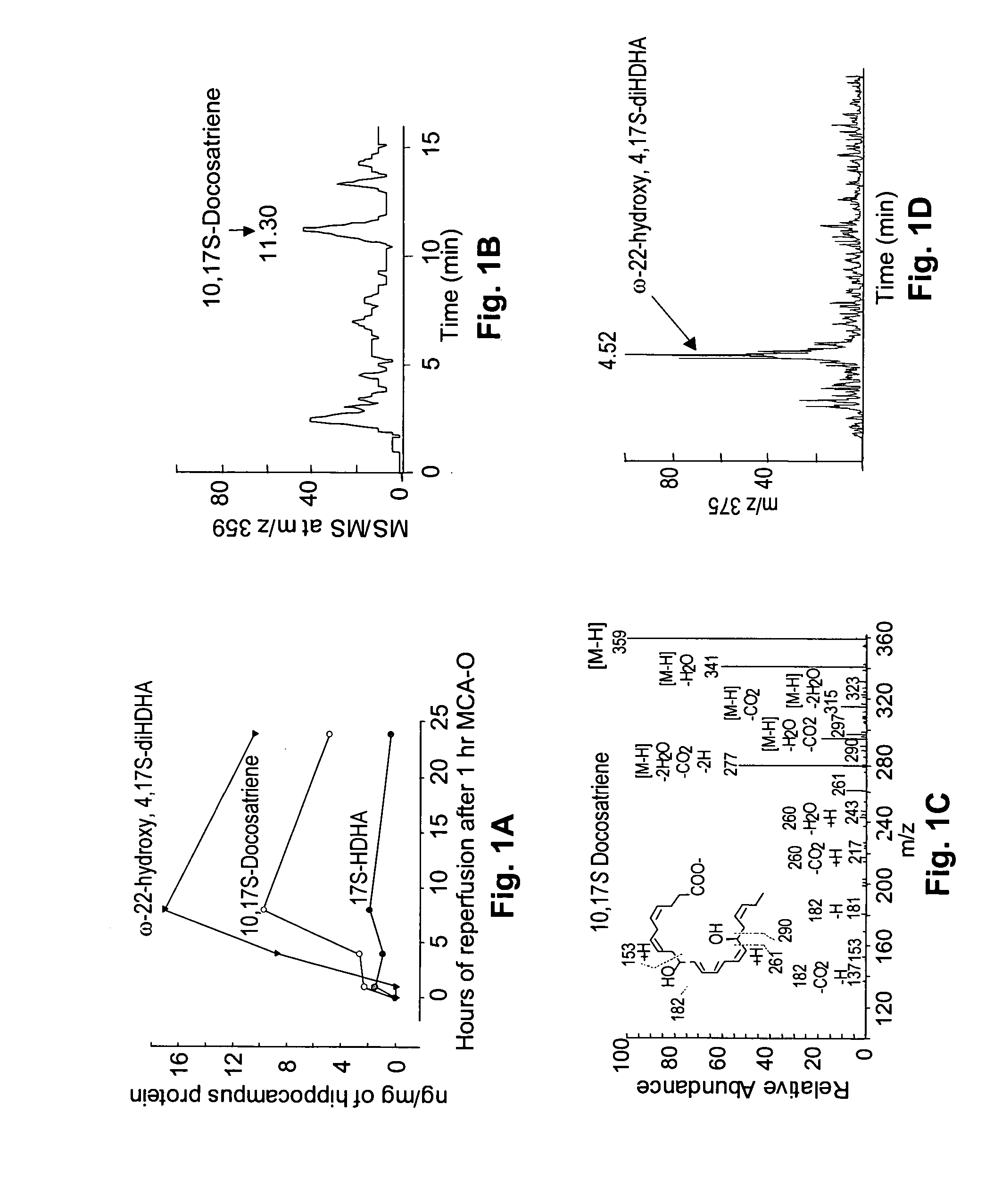
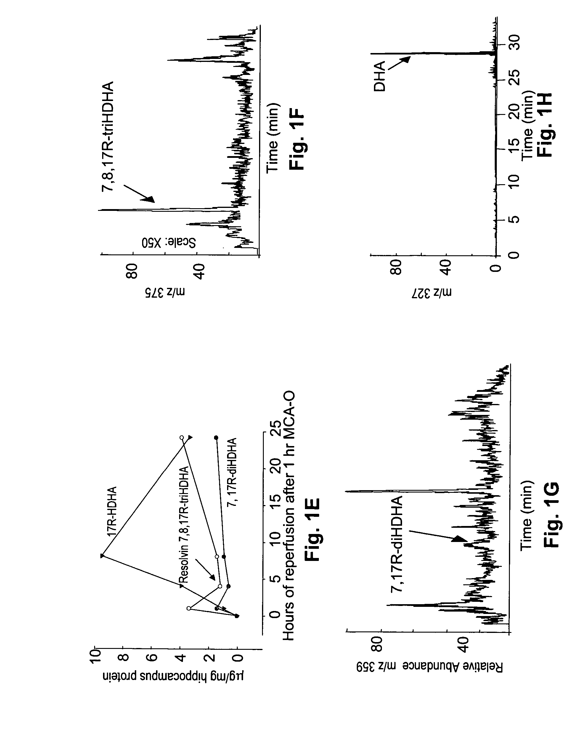
[0081] Right middle cerebral artery occlusion in mice for 1 h followed by reperfusion was used to assess the formation of docosahexaenoic acid (DHA)-oxygenation derivatives. Under these conditions there is active release of free docosahexaenoic acid from brain membrane phospholipids. This model of transient focal ischemia greatly affects the hippocampus, a brain region rich in neurons vulnerable in ischemic stroke, and in other neurologic diseases as described in Bazan, N. G., and Allan, G. (1998) In Cerebrovascular Disease: Pathophysiology, Diagnosis, and Management, eds. Ginsberg, M. D. and Bogousslavsky, J. (Blackwell Science, Inc., Maiden, Mass.) pp. 532-555.

[0082]FIGS. 1A-1H indicate the synthesis and metabolism of docosanoids in the ipsilateral mouse hippocampus during reperfusion following transient ischemia. The structural elucidation of DHA and docosanoids was performed by lipid...

example 3

Polymorphonuclear Leukocyte Infiltration Mediated by Focal Ischemic Stroke in Mice is Inhibited by 10,17S-docosatriene (NPD1)

[0093] Polymorphonuclear leukocyte (PMN) infiltration, a major factor in mediating brain ischemia-reperfusion damage was monitored. PMN infiltration is a complex, multi-step process that is modulated by the coordinated expression of adhesion and signaling molecules. DHA-derived messengers were very recently reported to inhibit PMN invasion outside the central nervous system, in the air-pouch model. (Hong et al., 2003).

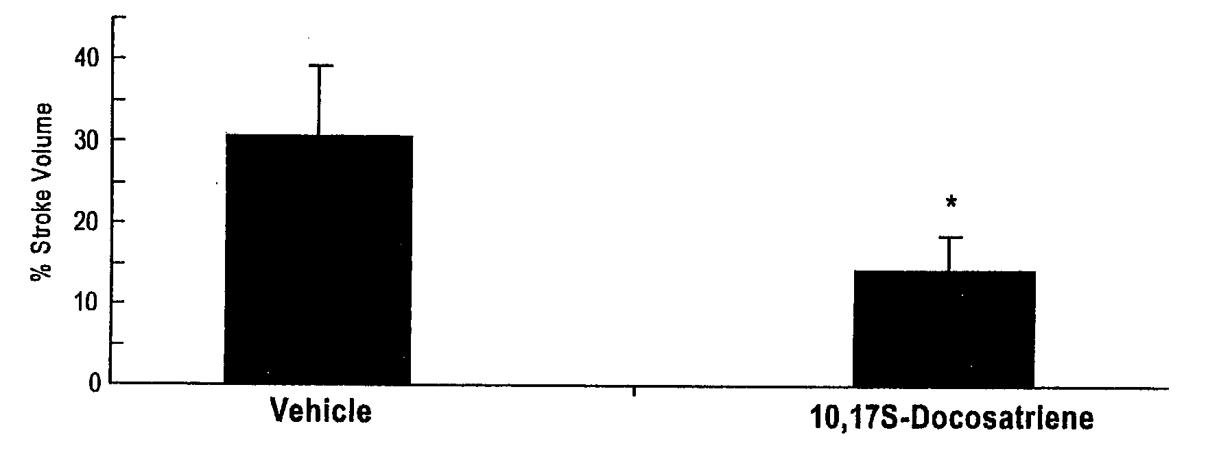
[0094] To determine whether 10,17S-docosatriene (NPD1) affected brain ischemia-reperfusion-induced PMN infiltration, either DHA or 10,17S-docosatriene (NPD1) was constantly infused into the third ventricle of a mouse brain during 48 h of reperfusion. FIGS. 5A, 5B, and 5C show the inhibition of leukocyte infiltration by 10,17S-docosatriene (NPD1) in mouse hippocampus and neocortex after 1-h MCA-O and 48 h of reperfusion. The data shown represent...


PUM


Abstract

A unique DHA product, 10, 17S-docosatriene (“Neuroprotectin D1” or “NPD1”), was found to provide surprisingly effective neuroprotection when administered right after an experimental stroke. Moreover, both nerve cells and retinal pigment epithelial (RPE) cells were found to synthesize 10,17S-docosatriene (NPD1) from DHA. NPD1 also potently counteracted H2O2/TNFα oxidative stress-mediated cell apoptotic damage. Under the same oxidative-stress conditions, NPD1 up-regulated the anti-apoptotic Bcl-2 proteins, Bcl-2 and Bcl-xL, and decreased expression of the pro-apoptotic proteins, Bad and Bax. Moreover, in RPE cells NPD1 inhibited oxidative stress-induced caspase-3 activation, IL-1β-stimulated human COX-2 promoter expression, and apoptosis due to N-retinylidene-N-retinylethanolamine (A2E). Overall, NPD1 protected both nerve and retinal pigment epithelial cells from cellular apoptosis and damage due to oxidative stress. NPD1 concentration in the brain of Alzheimer's patients was found to be significantly decreased from that of controls. In cultured human brain cells, NPD1 synthesis was up-regulated by neuroprotective soluble β amyloid, and NPD1 was found to inhibit secretion of toxic β amyloid peptides.

Description

[0001] The benefit of the filing dates of provisional application 60 / 493,110 filed 5 Aug. 2003, 60 / 564,426 filed 22 Apr. 2004, and 60 / 589,445 filed 20 Jul. 2004 are claimed under 35 U.S.C. § 119(e) in the United States, and are claimed under applicable treaties and conventions in all countries.[0002] This work was supported by National Institutes of Health grant nos. EY05121, NS23002, P20RR16816 from the COBRE Program, P01DE13499, and GM38765. The Government has certain rights in this technology.TECHNICAL FIELD [0003] This invention pertains to the use of 10,17S-docosatriene (“neuroprotectin D1” or “NPD1”), a product derived from docosahexaenoic acid (DHA), to protect cells from apoptosis, to protect the brain from damage due to ischemic stroke, to help prevent Alzheimer's Disease, and to help prevent retinal degeneration. BACKGROUND ART [0004] Docosanoids [0005] Dietary omega-3 fatty acids are required to maintain cellular functional integrity, and overall are necessary to human he...

Claims


Application Information

Patent Timeline
Patent Type & Authority Applications(United States)
IPC IPC(8): A61KA61K31/202
CPCA61K31/202A61P25/00A61P25/28A61P27/02
Inventor BAZAN, NICOLAS G.SERHAN, CHARLES N.MARCHESELLI, VICTOR L.MUKHERJEE, PRANAB K.BARREIRO, SEBASTIAN G.LUKIW, WALTER J.HONG, SONGGRONERT, KARSTENMUSTO, ALBERTO E.
Owner THE BRIGHAM & WOMEN S HOSPITAL INC
Why Eureka
Try Eureka