Peptides that inhibit complement activation
- Summary
- Abstract
- Description
- Claims
- Application Information
AI Technical Summary
Benefits of technology
Problems solved by technology
Method used
Image
Examples
example 1
Panning Against C5
[0150] Peptides were identified by panning an M13 phage library (New England Biolabs), a random 12mer peptide library with a diversity of 1011 different peptide structures, against complement component C5. The phage library was panned against C5 using three different methods for immobilizing C5. The binding reactions all contained 2×10¹¹ phage particles in PBS containing 0.5% BSA and 0.3% Tween 20. The binding reactions were carried out at 25° C. for two hours and then unbound phage were removed by washing 5 times with PBS containing 0.5% BSA and 0.3% Tween20. The phage were eluted by acid (0.2 M Glycine pH 2.2, 1 mg / mL BSA) and immediately neutralized with 1 M Tris pH 9.1. Following elution, recovered phage were amplified in ER2738 E. coli (NEB) and subjected to two more rounds of panning as described above. The methods for immobilizing C5 include directly coating C5 (Advanced Research Technologies) to the surface (1 μg / mL), capturing biotinylated C5 (1 μg / mL) on...
example 2
Phage Clone Binding to C5
[0153] The binding sites of the peptides were further probed via C5-binding competition assays. Increasing concentrations of each phage clone were incubated for 2 hours with 200 ng of biotinylated C5 in PBS containing 0.5% BSA. The phage bound C5 complex was captured on a neutravidin coated microplate for 20 minutes at room temperature and then washed with PBS containing 0.5% BSA. A peroxidase labeled anti-M13 antibody (Pharmacia) was used to detect the amount of phage bound to C5 with OPD substrate. Competition assays with free peptide were carried out in a similar manner except that one concentration of each phage clone was used which was determined as the concentration giving approximately 50% binding in the previous titration. Free peptide was added to the phage clones at concentrations of 50, 10 and 1 μM and incubated with the biotinylated C5 and the assay was carried out as above.
[0154] The binding assays showed all peptides bound C5. Furthermore, th...
example 3
Classical Pathway Hemolytic Assay
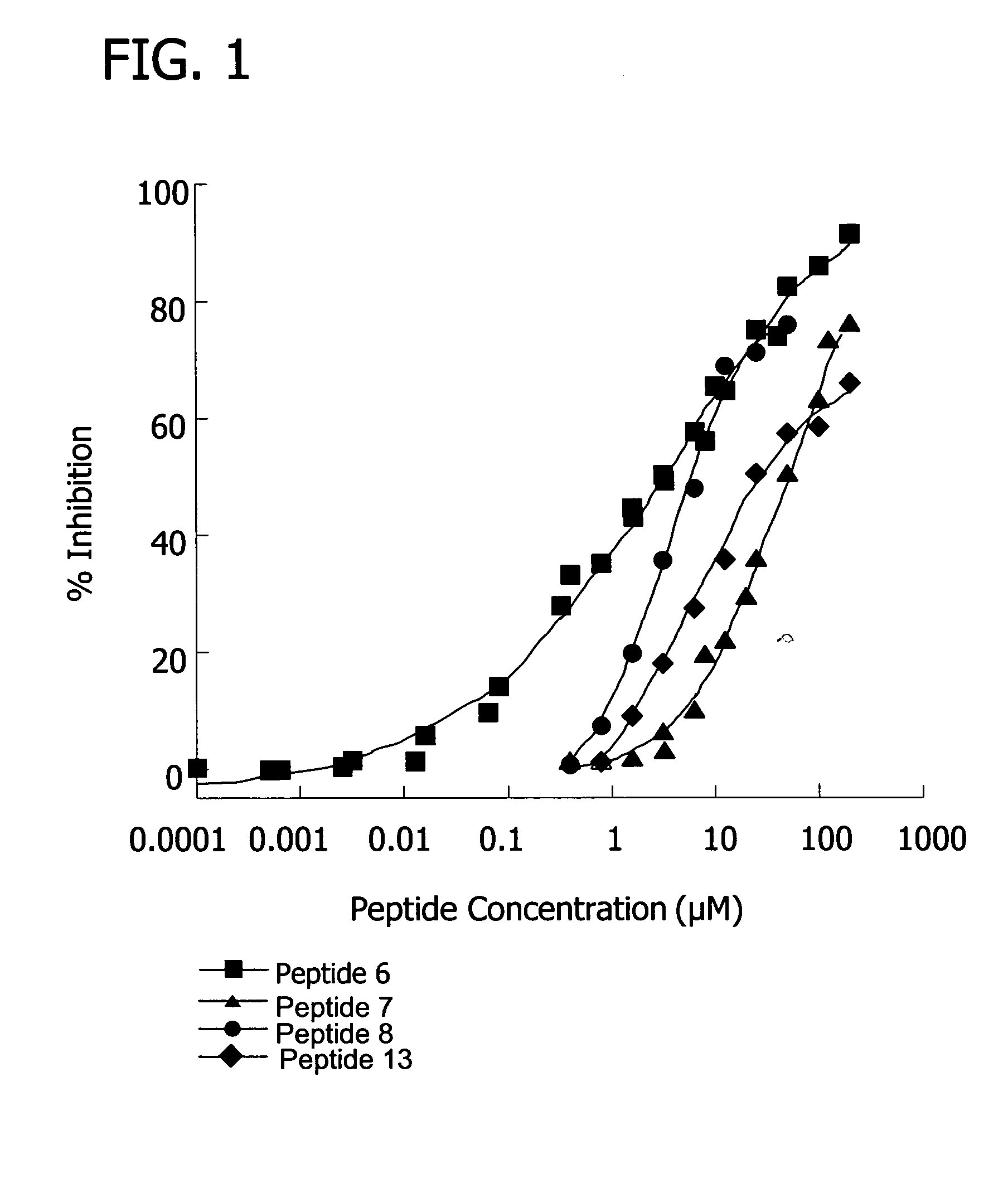
[0155] Inhibition of the classical pathway by the peptides was measured using a standard hemolysis assay, which monitors the lysis of sheep erythrocytes as a result of terminal complex formation (see e.g. FIG. 1, FIG. 10). Various concentrations of peptide were incubated with 50 μL pooled human plasma (diluted 1:30 with GVB++), 5×10⁷ antibody sensitized sheep erythrocyte cells, and GVB++ buffer to a final volume of 200 μL. The reaction was incubated at 37° C. for one hour and centrifuged. The percentage of lysis was determined by measuring the optical density of the supernatant at 414 nm. 100% lysis was determined by the optical density at 414 nm of the supernatant from a control sample consisting of 5×10⁷ antibody sensitized sheep erythrocyte cells in water. The percent inhibition was calculated as the (% lysisno peptide−% lysispeptide) / % lysisno peptide×100.
[0156] Results showed that, at 100 uM concentrations, several peptides (peptide 7, SEQ ID ...
PUM
| Property | Measurement | Unit |
|---|---|---|
| Composition | aaaaa | aaaaa |
| Polarity | aaaaa | aaaaa |
| Acidity | aaaaa | aaaaa |
Abstract
Description
Claims
Application Information
 - R&D
- Intellectual Property
- Life Sciences
- Materials
- Tech Scout
- Unparalleled Data Quality
- Higher Quality Content
- 60% Fewer Hallucinations
Browse by: Latest US Patents, China's latest patents, Technical Efficacy Thesaurus, Application Domain, Technology Topic, Popular Technical Reports.
© 2025 PatSnap. All rights reserved.Legal|Privacy policy|Modern Slavery Act Transparency Statement|Sitemap|About US| Contact US: help@patsnap.com


