Devices for applying energy to tissue

a tissue and energy technology, applied in the field of tissue energy devices, can solve the problems of copd-related injuries, inability to perform common daily activities, and loss of muscle strength of individuals, and achieve the effects of reducing the risk of copd, and reducing the chance of recovery

Inactive Publication Date: 2005-05-19
BRONCUS MEDICAL
View PDF99 Cites 180 Cited by
  • Summary
  • Abstract
  • Description
  • Claims
  • Application Information

AI Technical Summary

Problems solved by technology

Those inflicted with COPD face disabilities due to the limited pulmonary functions.
Usually, individuals afflicted by COPD also face loss in muscle strength and an inability to perform common daily activities.
Since the damage to the lungs is irreversible, there is little hope of recovery.
Most times, the physician cannot reverse the effects of the disease but can only offer treatment and advice to halt the progression of the disease.
However, these conducting airways do not take part in gas exchange because they do not contain alveoli.
If the lungs' ability to recoil is damaged, the lungs cannot contract and reduce in size from their inflated state.
As a result, the lungs cannot evacuate all of the inspired air.
The destruction of the alveolar walls results in a dual problem of reduction of elastic recoil and the loss of tethering of the airways.
Unfortunately for the individual suffering from emphysema, these two problems combine to result in extreme hyperinflation (air trapping) of the lung and an inability of the person to exhale.
In this situation, the individual will be debilitated since the lungs are unable to perform gas exchange at a satisfactory rate.
While alveolar wall destruction decreases resistance to collateral ventilation, the resulting increased collateral ventilation does not benefit the individual since air is still unable to flow into and out of the lungs.
Yet, bronchodilator drugs are only effective for a short period of time and require repeated application.
Moreover, the bronchodilator drugs are only effective in a certain percentage of the population of those diagnosed with COPD.
Unfortunately, aside from the impracticalities of needing to maintain and transport a source of oxygen for everyday activities, the oxygen is only partially functional and does not eliminate the effects of the COPD.
Moreover, patients requiring a supplemental source of oxygen are usually never able to return to functioning without the oxygen.
However, lung reduction surgery is an extremely traumatic procedure which involves opening the chest and thoracic cavity to remove a portion of the lung.
If the entire lung is emphysematous, however, removal of a portion of the lung removes gas exchanging alveolar surfaces, reducing the overall efficiency of the lung.
Lung volume reduction surgery is thus not a practical solution for treatment of emphysema where the entire lung is diseased.
Both bronchodilator drugs and lung reduction surgery fail to capitalize on the increased collateral ventilation taking place in the diseased lung.
Such a need is evident in dynamically moving environments (e.g., the lungs) where repositioning of a device to find the original target site may be difficult.
However, since the characteristics of components used to detect a Doppler shift vary from characteristics of components used to cut or remove tissue, it is difficult to cut or remove tissue in precisely the same location and immediately after detection has taken place.
For instance, if a device uses energy to create an opening or ablate tissue, the energy delivery components may not have acceptable characteristics to function as Doppler components.
Furthermore, the process of delivering energy through the device may undesirably impact any Doppler components.
As a result, the ultrasound signal may experience significant reflection and divergence at the tissue / transducer interface.
One drawback to using Doppler ultrasound devices for placing collateral openings in tissue is that conventional tip materials selected for their desirable acoustic impedance are not effective to deliver energy (e.g., RF, resistive heat, etc.) The acoustic impedance of electrically and thermally conductive materials is higher than the desired acoustic impedance of 6.79 MRayls.
Another drawback to delivering energy through devices configured for Doppler applications is that the transducer is prone to being damaged.
Moreover, conduction of heat through the device may adversely affect the joints and bonds between the transducer, tip and device.
As a result, there is the potential of a catastrophic failure of the device if the assembly breaks apart during use in the body.

Method used


Image

  • Devices for applying energy to tissue
  • Devices for applying energy to tissue
  • Devices for applying energy to tissue

Examples


Embodiment Construction

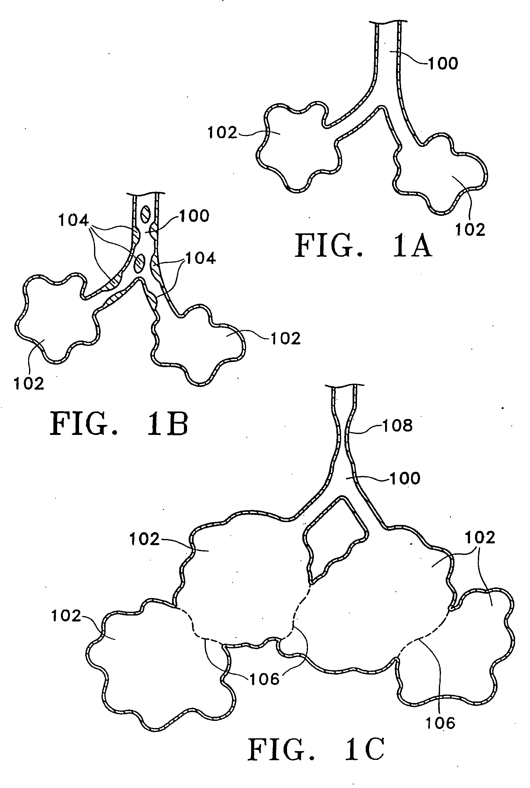
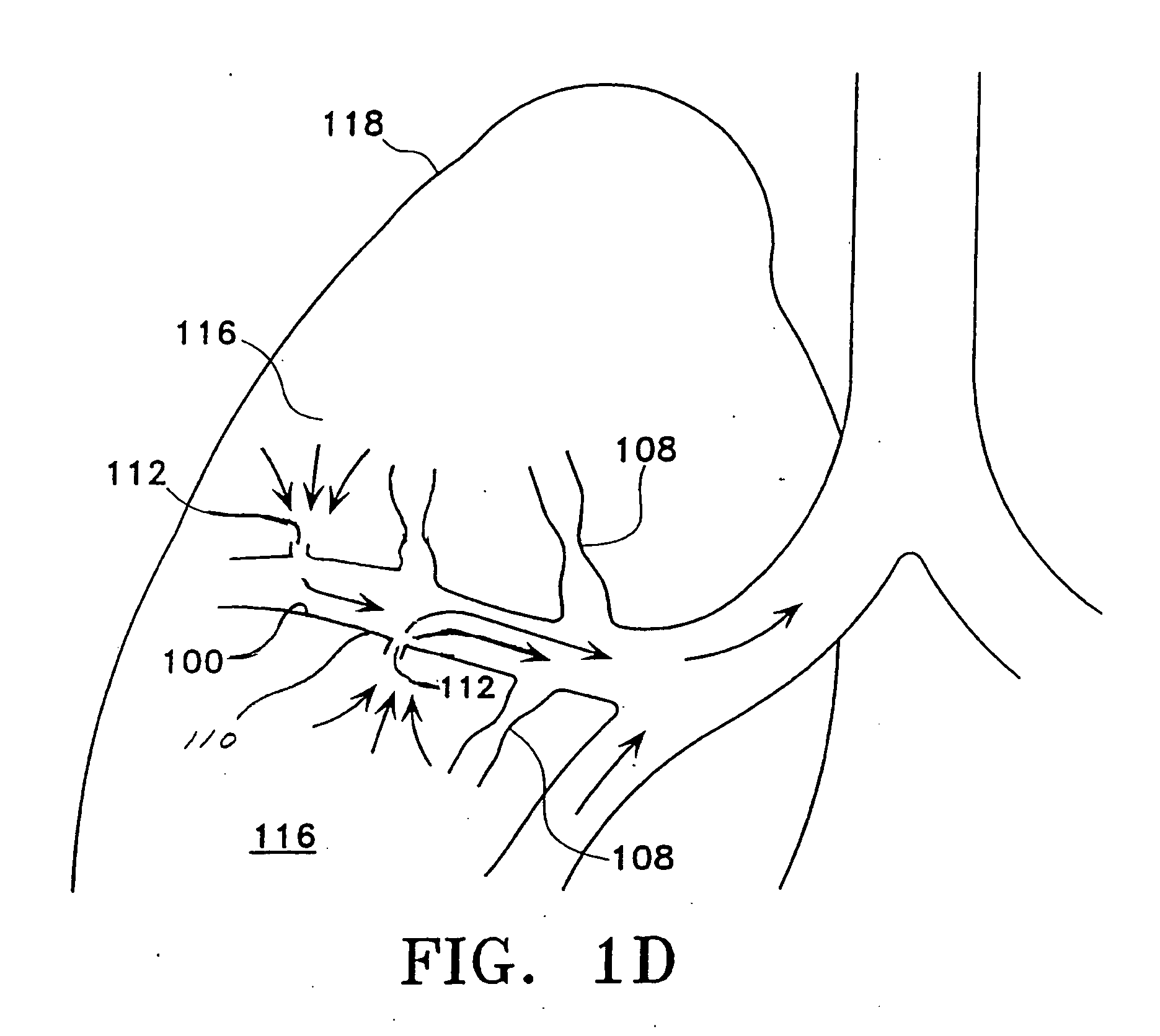
[0055] Prior to considering the invention, simplified illustrations of various states of a natural airway and a blood gas interface found at a distal end of those airways are provided in FIGS. 1A-1C. FIG. 1A shows a natural airway 100 which eventually branches to a blood gas interface 102. FIG. 1B illustrates an airway 100 and blood gas interface 102 in an individual having COPD. The obstructions 104 (e.g., excessive mucus resulting from COPD, see above) impair the passage of gas between the airways 100 and the interface 102. FIG. 1C illustrates a portion of an emphysematous lung where the blood gas interface 102 expands due to the loss of the interface walls 106 which have deteriorated due to a bio-chemical breakdown of the walls 106. Also depicted is a constriction 108 of the airway 100. A combination of the phenomena depicted in FIGS. 1A-1C are often found in the same lung.

[0056] The following text and corresponding figures provide variations and embodiments of the present inven...


PUM


Abstract

Disclosed herein are devices for altering gaseous flow within a lung to improve the expiration cycle of an individual, particularly individuals having chronic obstructive pulmonary disease (COPD). More particularly, a medical catheter is disclosed to detect the presence of blood vessels and to produce collateral openings or channels through the airway wall so that air is able to pass directly out of the lung tissue to facilitate both the exchange of oxygen ultimately into the blood and/or to decompress hyper-inflated lungs.

Description

CROSS REFERENCE TO RELATED APPLICATIONS [0001] This application is a continuation of U.S. application Ser. No. 10 / 280,851 filed Oct. 25, 2002, which is a continuation in part of U.S. application Ser. No. 10 / 080,344, filed Feb. 21, 2002, which is a continuation in part of U.S. application Ser. No. 09 / 946,706, filed Sep. 4, 2001, which claims the benefit of U.S. Provisional Application No. 60 / 269,130, filed on Feb. 14, 2001. U.S. application Ser. No. 09 / 946,706, filed Sep. 4, 2001, is a continuation in part of U.S. application Ser. No. 09 / 633,651, filed Aug. 7, 2000, which claims the benefit of U.S. Provisional Application No. 60 / 147,528, filed on Aug. 5, 1999, and U.S. Provisional Application No. 60 / 176,141, filed on Jan. 14, 2000. Each of the above referenced applications is incorporated herein by reference in its entirety.FIELD OF THE INVENTION [0002] The invention is directed to devices for altering gaseous flow within a lung to improve the expiration cycle of an individual, parti...

Claims


Application Information

Patent Timeline
Patent Type & Authority Applications(United States)
IPC IPC(8): A61BA61B8/06A61B8/12A61B17/00A61B17/064A61B17/08A61B17/11A61B17/22A61B17/32A61B18/00A61B18/14A61B18/18A61B19/00A61F2/02A61F2/04A61F2/06A61F2/20A61F2/24
CPCA61B5/489A61B8/445A61B8/12A61B17/064A61B17/0644A61B17/08A61B17/11A61B17/22A61B17/320068A61B18/1477A61B18/1485A61B18/1492A61B18/1815A61B19/52A61B2017/00106A61B2017/00252A61B2017/00575A61B2017/1135A61B2017/1139A61B2017/22067A61B2017/22077A61B2018/00005A61B2018/00029A61B2018/00214A61B2018/00273A61B2018/00285A61B2018/00541A61B2018/00601A61B2018/00982A61B2018/1425A61B2018/1437A61B2018/1475A61B2019/481A61B2019/5278A61B2019/528A61B2019/545A61F2/02A61F2/20A61F2/2412A61F2/91A61F2002/043A61F2002/061A61F2002/8483A61N2007/0078A61B8/06A61B90/36A61B2090/3782A61B2090/3784A61B2090/08021A61B2090/395A61B2017/320069
Inventor TOM, CURTIS P.KRAMER, THOMAS A.ROSCHAK, EDTANAKA, DONTHOMPSON, DAVID
Owner BRONCUS MEDICAL
Why Eureka
Try Eureka