Method and assembly for distal embolic protection

a technology of distal embolic protection and assembly, which is applied in the field of cardiovascular surgery, can solve the problems of long and tedious open-heart valve repair or replacement surgery, prolonged hospitalization and painful recovery period for patients, and significant trauma, and achieve the effect of reducing the retrograde flow

Active Publication Date: 2005-06-02
MEDTRONIC 3F THERAPEUTICS
View PDF40 Cites 284 Cited by
  • Summary
  • Abstract
  • Description
  • Claims
  • Application Information

AI Technical Summary

Benefits of technology

[0021] The filter assemblies disclosed herein may further comprise a cloth covering the perimeter of the open proximal end of the porous bag. Used in this manner, the cloth covering will substantially form a seal between the open proximal end and the walls of the blood vessel. Such a seal will ensure that embolic material and debris will not be trapped in or be allowed to pass between the open proximal end of the porous bag and the walls of the blood vessel. In addition, the cloth covering will protect the aortic wall from becoming damaged by the expanding frame of the filter assembly.
[0022] Additionally and alternatively, the filter assemblies disclosed herein may further include a one-way valve at the open proximal end of the filter assembly to serve the dual function of acting as a temporary valve during valve replacement surgery and preventing embolic material and debris from escaping out from the filter. The valve permits the natural forward flow of b

Problems solved by technology

Open-heart valve repair or replacement surgery is a long and tedious procedure and involves a gross thoracotomy, usually in the form of a median sternotomy.
This method, however, is highly invasive and often results in significant trauma, risk of complications, as well as extended hospitalization and painful recovery period for the patient.
The use of a minimally invasive approach, however, introduces new complexities to surgery.
An inherent difficulty in the minimally invasive percutaneous approach is the limited space that is available within the vasculature.
Consequently, the introduction of tools and prosthetic devices becomes a great deal more complicated.
Accordingly, while heart valve surgery produces beneficial results for many patients, numerous others who might benefit from such surgery are either unable or unwilling

Method used


Image

  • Method and assembly for distal embolic protection
  • Method and assembly for distal embolic protection
  • Method and assembly for distal embolic protection

Examples


Embodiment Construction

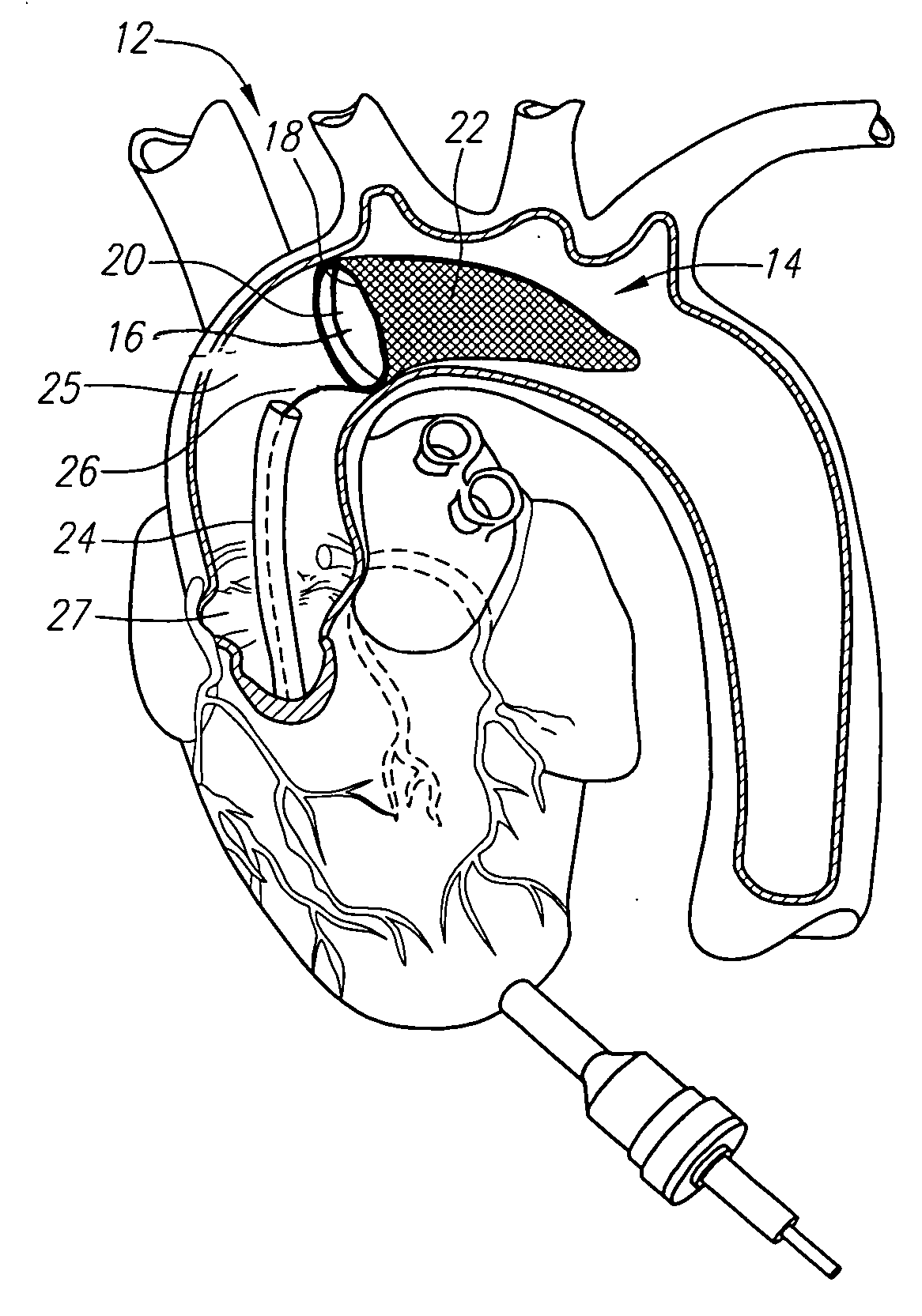
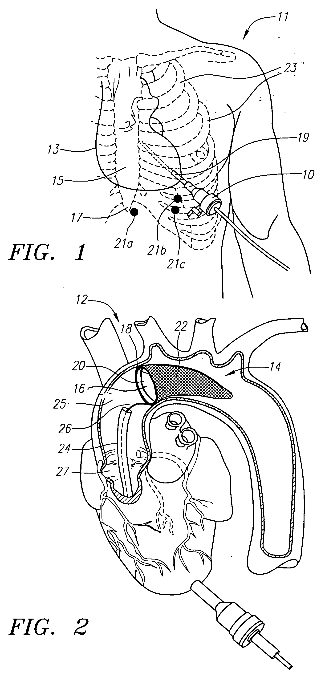
[0033] The advantages of performing valve repair or replacement surgery through the apical area of a patient's heart have been described in co-pending patent application Ser. No. 10 / 831,770, filed Apr. 23, 2004, which is incorporated herein by reference. The apical approach is significantly less invasive than open-chest techniques and it provides a more direct surgical approach to the valves and great vessels of the heart than the conventional minimally invasive percutaneous techniques. Moreover, because the apical approach can accommodate a larger incision and does not require maneuvering through long convolutions of the vasculature from the femoral arteries, it is not limited by the size constraints of the percutaneous techniques. Moreover, percutaneous methods may not be suitable in patients with severe atherosclerosis in which the vasculature is substantially narrowed.

[0034] The apical approach to valve replacement surgery is particularly suited for replacement of heart valves,...


PUM


Abstract

Methods and assemblies are described for capturing embolic material in a blood vessel or other body cavity during cardiovascular or valve replacement and repair surgery, wherein access is provided through the apical area of the patient's heart. The distal embolic protection assembly generally comprises a sleeve having a lumen, an actuating member having proximal and distal ends, wherein the actuating member is movably disposed within the lumen, and a filter assembly coupled to the distal end of the actuating member. The filter assembly generally comprises a porous bag having an open proximal end, a collapsible and expandable frame that is coupled to the open proximal end of the porous bag, and at least one support spine disposed at least a part of the longitudinal axis of the porous bag. The porous bag is configured such that it permits blood to perfuse freely through while capturing embolic material and other debris.

Description

CROSS-REFERENCE TO RELATED APPLICATION [0001] This application is a continuation-in-part of, and claims priority to, co-pending patent application Ser. No. 10 / 680,717, filed Oct. 6, 2003 and co-pending patent application Ser. No. 10 / 831,770, filed Apr. 23, 2004, the entirety of which is incorporated by reference in their entirety.FIELD OF THE INVENTION [0002] The present invention relates generally to methods and systems for cardiovascular surgery. More particularly, the invention relates to methods and systems for capturing embolic or other materials during cardiovascular surgery. BACKGROUND OF THE INVENTION [0003] Various surgical techniques may be used to repair a diseased or damaged heart valve, such as annuloplasty (contracting the valve annulus), quadrangular resection (narrowing the valve leaflets), commissurotomy (cutting the valve commissures to separate the valve leaflets), or decalcification of valve and annulus tissue. Alternatively, the diseased heart valve may be repla...

Claims


Application Information

Patent Timeline
IPC IPC(8): A61M29/00
CPCA61F2/013A61F2/24A61F2250/0003A61F2230/0067A61F2230/0006A61F2230/0008A61F2002/018A61F2/014A61F2/2476
Inventor BERGHEIM, BJARNE
Owner MEDTRONIC 3F THERAPEUTICS
Why Eureka
Try Eureka