Articular cartilage repair implant delivery device and method of use

a technology for articular cartilage and implants, which is applied in the field of articular cartilage repair implants delivery devices and methods of use, can solve the problems of limited spontaneous healing of damaged articular cartilage, increased risk of injury to cartilage, and articular cartilage tears

Inactive Publication Date: 2005-06-23
DEPUY PROD INC
View PDF20 Cites 309 Cited by
  • Summary
  • Abstract
  • Description
  • Claims
  • Application Information

AI Technical Summary

Benefits of technology

[0027] According to another aspect of the principles of the present invention, there is provided a set of surgical tools for preparing a damaged articular cartilage site on subchondral bone of a joint and implanting an articular cartilage repair device. The device includes a cannula, surgical drill and implant delivery device. The cannula has a tubular body defining a proximal and a distal end a bore disposed in the tubular body and extending from the proximal end to the distal end, a blade disposed at a tip of the distal end of the tubular body and configured to incise about an articular cartilage area. The surgical drill has a drill shaft having a proximal end and a distal end, an attachment tip on the proximal end of the drill shaft and configured to be received in a rotation device, and a site preparation tip on the distal end of the dri

Problems solved by technology

In addition, as a person ages, articular cartilage loses mechanical strength, rendering the cartilage even more susceptible to trauma, such that even common motions (e.g. squatting, stair climbing, etc.) can cause articular cartilage tears.
Because articular cartilage tissue is aneural (little or no nerves) and avascular (little or no blood vessels), the spontaneous healing of damaged articular cartilage is limited.
As a result, focal (localized) defects can tend to lead toward progressive degeneration of the joint surface(s) until total joint replacement is eventually necessary.
In addition, it is generally agreed that while this method is able to produce short-term alleviatio

Method used


Image

  • Articular cartilage repair implant delivery device and method of use
  • Articular cartilage repair implant delivery device and method of use
  • Articular cartilage repair implant delivery device and method of use

Examples


Embodiment Construction

[0068] While the invention is susceptible to various modifications and alternative forms, specific embodiments thereof have been shown by way of example in the drawings and will herein be described in detail. It should be understood, however, that there is no intent to limit the invention to the particular forms disclosed, but on the contrary, the intention is to cover all modifications, equivalents and alternatives falling within the spirit and scope of the invention as defined by the appended claims.

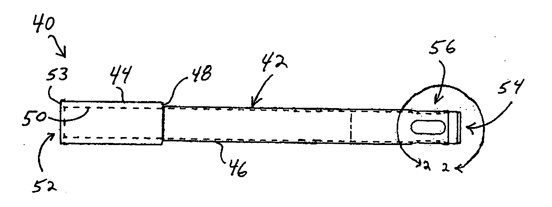
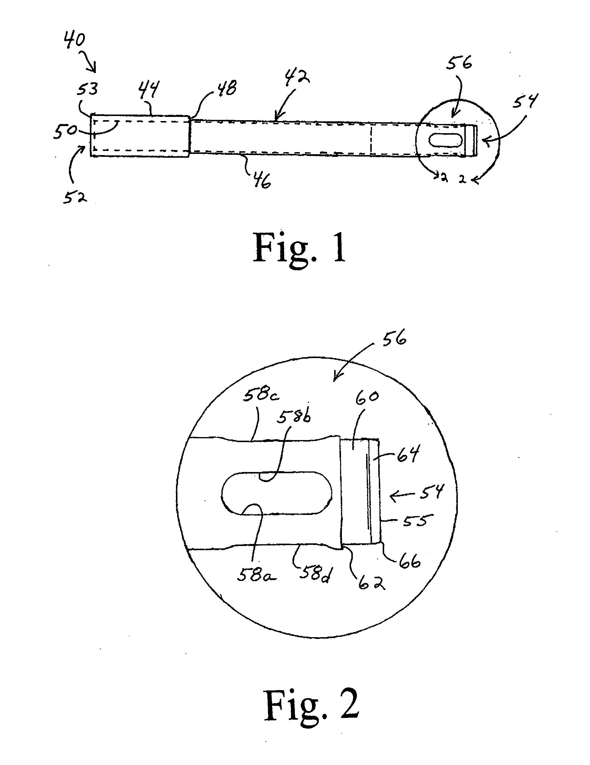
[0069] Referring now to FIG. 1, there is depicted an exemplary embodiment of one form of a cannula or cannula construct, generally designated 40, fashioned in accordance with an aspect of the principles of the subject invention. While the cannula 40 is an instrument unto itself, the cannula 40 is one of a set of instruments or devices for performing a surgical procedure on articular cartilage, the other instruments of the set being shown in the other figures and described herein. Whil...


PUM


Abstract

A technique for the arthroscopic delivery and fixation of an articular cartilage repair device or implant is provided. The technique includes the use of a cannula tube that functions as both a cartilage cutter and a guide to pass instruments into the body arthroscopically. One such instrument is an end-cutting reamer that both prepares the subchondral bone by re-surfacing it down to a specified depth and also simultaneously drills a pilot hole in the subchondral bone to accept the cartilage repair device. A delivery device is utilized to hold and deliver the cartilage repair device to the delivery site.

Description

FIELD OF THE INVENTION [0001] The present invention relates to repair of articular cartilage in joints and, more particularly, to a technique and instruments for preparing subchondral tissue and bone, and implanting an articular cartilage repair unit therein. BACKGROUND OF THE INVENTION [0002] Articular cartilage is a type of hyaline cartilage that lines the surfaces of the opposing bones in a diarthrodial joint (e.g., knee, hip, shoulder, etc.). Its primary function is to permit smooth, near frictionless movement during articulation between bones of the joint by providing a low-friction interface between the contacting cartilage surfaces of the joint. Articular cartilage is also load bearing, and serves to transmit and distribute compressive joint loads to the underlying subchondral bone (i.e. bone beneath the cartilage or subchondral tissue). [0003] Articular cartilage is typically damaged in one of two ways, acute trauma inflicted through physical activity (such as twisting motio...

Claims


Application Information

Patent Timeline
IPC IPC(8): A61B17/00A61B17/16A61B17/32A61B17/34A61B19/00A61F2/30A61F2/46
CPCA61B17/00234A61F2002/4627A61B17/1631A61B17/1635A61B17/1637A61B17/320016A61B17/320036A61B17/3421A61B2017/1602A61B2017/3445A61B2019/461A61B2019/462A61F2/30749A61F2/30756A61F2/4618A61F2002/30751A61F2002/30766A61F2002/30878A61F2002/4635A61F2002/4661A61F2002/4662A61B17/1615A61B2090/061A61B2090/062
Inventor JACOBS, ANDREW M.ZANNIS, ANTHONY D.SCHWARTZ, HERBERT E.DAY, CAROLYN K.KEMPPAINEN, JOHN
Owner DEPUY PROD INC
Why Eureka
Try Eureka