
Cystotomy catheter capture device and methods of using same

a technology of capturing device and catheter, which is applied in the field of medical devices, can solve the problems of inability to void satisfactorily, complicating postoperative care, and many women, however, are either unable to learn or do not want to place a catheter blindly

Inactive Publication Date: 2005-06-30
SWAN VALLEY MEDICAL
View PDF14 Cites 110 Cited by
  • Summary
  • Abstract
  • Description
  • Claims
  • Application Information

AI Technical Summary

Benefits of technology

[0017] A first embodiment of the catheter capture device of the present invention comprises a urethral sound and a sleeve, wherein the urethral sound is inserted through a patient's urethra into the bladder and out the abdominal wall, the sound comprises a removable tip, the removable tip of the sound is removed, the sleeve is attached to the end of the sound from which the removable tip was removed, the catheter to be captured comprises a balloon and a tip, the catheter tip is inserted into the sleeve, the balloon is inflated, and the catheter is pulled into the patient's bladder by pulling the sound out through the patient's urethra. The sleeve optionally comprises a flange that prevents the catheter from falling out of the sleeve when the balloon is inflated.
[0025] The present invention also covers a number of different methods of capturing a catheter, corresponding to the catheter capture devices described above. The first method comprises the steps of: inserting a urethral sound through a patient's urethra into the bladder and out the abdominal wall; wherein the urethral sound comprises a removable tip, removing the removable tip; attaching a sleeve to the end of the sound from which the removable tip was removed; wherein the catheter to be captured comprises a tip, inserting the catheter tip into the sleeve; wherein the catheter to be captured comprises a balloon, inflating the balloon; and pulling the catheter into the patient's bladder by pulling the sound out through the patient's urethra. The sleeve optionally comprises a flange that prevents the catheter from falling out of the sleeve when the balloon is inflated.

Problems solved by technology

A large percentage of these women are unable to void satisfactorily post-operatively and require a catheter to drain the bladder for several days or weeks.
PUR is generally only a temporary event lasting a few days to weeks, but it can be painful, frightening and distressing, and it can complicate postoperative care.
Many women, however, are either unable to learn or do not want to place a catheter blindly into the urethra and bladder, through a painful, freshly operated area with sutures that are oozing blood and serum (5).
Smaller catheters (i.e., catheters with a smaller diameter—not length) are used with the O / I techniques because O / I can cause perforation of the bowel or peritoneal cavity, and larger tubes (or catheters) would lead to a higher complication rate.
The I / O method, despite its advantages, has been awkward and difficult with current devices.
In comparison to I / O techniques, the O / I technique is simple, cheap and easy, but bladder drainage is unreliable because the small catheters often kink or become obstructed when small blood clots enter or form inside the catheter.
As a result, the O / I technique is never used for permanent catheterization because of unreliable urine drainage.
All O / I devices are more prone to unrecognized bowel or peritoneal perforation with serious secondary complications than the I / O devices.
The applicant believes that an important reason for the current popularity of O / I techniques is because the I / O devices that are currently available are poorly designed, awkward and difficult to use.
Moreover, catheter capture is difficult to achieve with these I / O devices.
Each of the devices currently on the market, however, has serious drawbacks.
One drawback that is common to all of these devices is that the tip of each device has a short “throw” so that it is difficult to pass the tip of the device through the abdominal wall.
When the device is too short to advance through the abdominal wall, catheter capture (i.e., securing or affixing the catheter) becomes extremely difficult.
Another drawback is that existing catheter capture methods do not work.
The Giesy device is limited in that it only describes two means of coupling the catheter to the placement device.
The Leach device is limited in that it has a short “throw” and uses a jaw mechanism to capture the catheter.
The jaw mechanism becomes wider after the catheter is enclosed within the jaws, making it more difficult for the catheter to be pulled safely through a small hole in the bladder and potentially resulting in loss of the catheter.
Loss of the catheter requires the surgeon to start all over again, subjecting the patient to further unnecessary trauma.
The Lyell device is limited in that the only catheter capture means it describes is a hook on the end of a flexible wire.
Because of the problems associated with current suprapubic catheter placement technologies, many patients have been placed on urethral catheterization immediately after surgery instead of suprapubic bladder catheterization during surgery.
Specifically, residual urine is impossible to determine while an indwelling urethral catheter is present to drain the bladder because the catheter fills the urethra and makes it impossible to void.
Patients are much more comfortable with suprapubic catheters than with urethral catheters exiting the genitalia, and sexual relations are impossible with a urethral catheter in place.
Thus, the preferred alternative is still I / O suprapubic catheter placement, but current methods and available devices are inadequate—particularly in cases involving women, where the distance from the bladder to the abdominal is often greater than in men.
Most of these patients do not have suprapubic catheterization because it would be a difficult and formidable procedure for them as currently performed.

Method used


Image

  • Cystotomy catheter capture device and methods of using same
  • Cystotomy catheter capture device and methods of using same
  • Cystotomy catheter capture device and methods of using same

Examples


Embodiment Construction

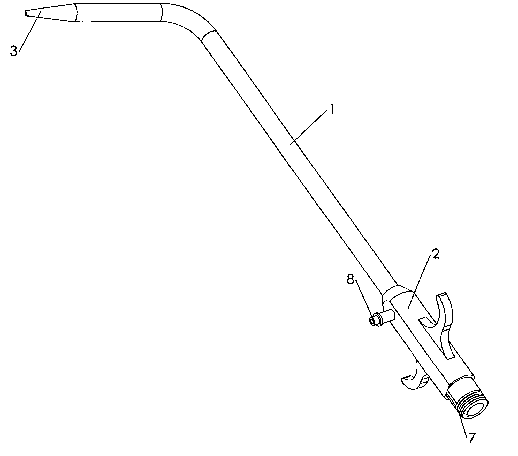
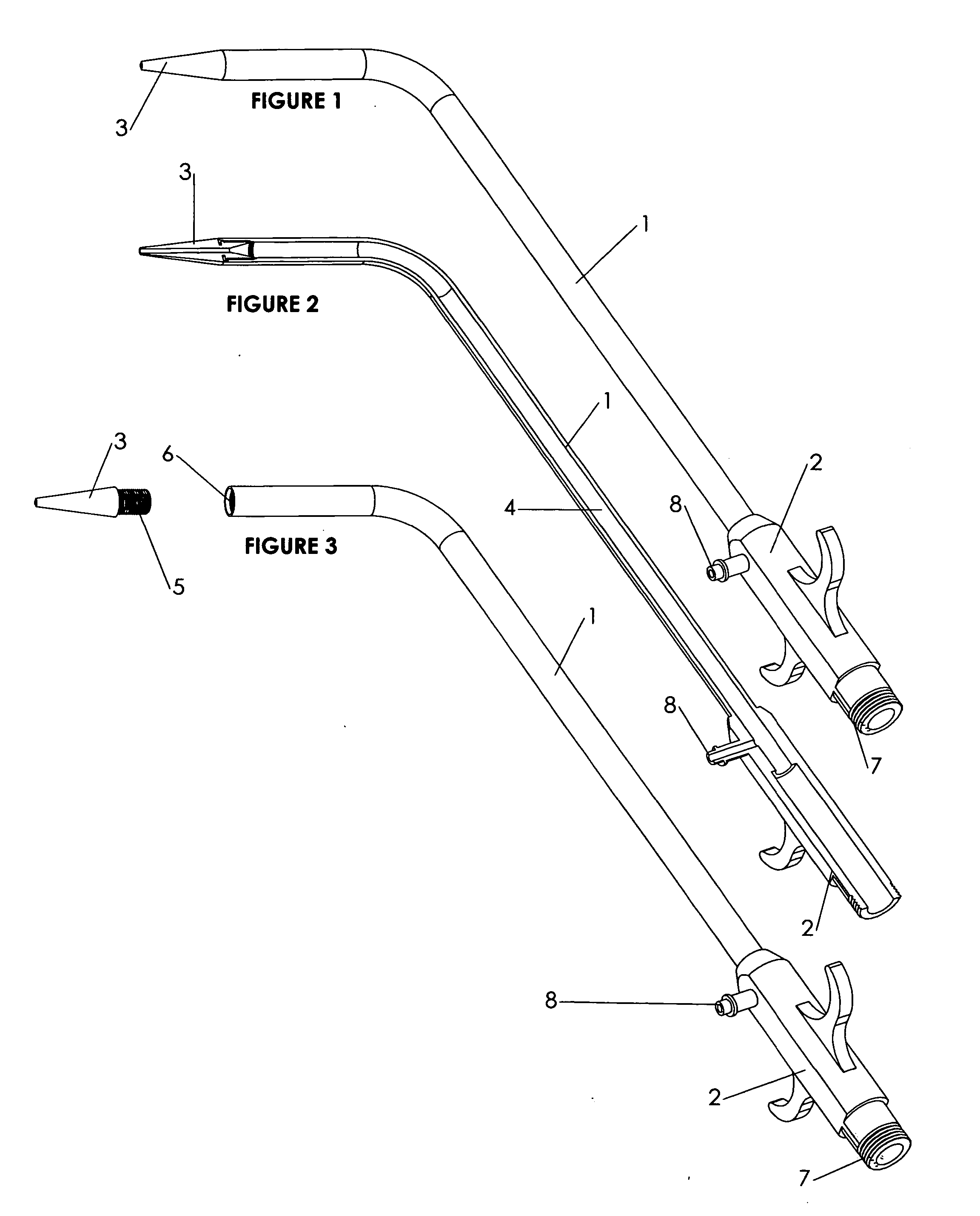
[0135]FIG. 1 is a perspective view of the urethral sound of the present invention. This figure shows the sound 1, the handle 2, and the tip 3. The sound is hollow, and there are holes ar either end of the sound for the insertion of a wire.

[0136]FIG. 2 is a section view of the urethral sound of the present invention. This figure shows the sound 1, the handle 2, the tip 3, and the hollow channel 4, which extends from one end of the sound to the other.

[0137]FIG. 3 is a perspective view of the urethral sound of the present invention with the tip removed. This figure shows the sound 1, the handle 2, and the tip 3. It also shows the threaded end 5 of the tip, which is inserted into the threaded distal end 6 of the sound. The proximal end of the sound 7 is also threaded for the addition of a Tuohy-Borst adapter or an endoscopic cap.

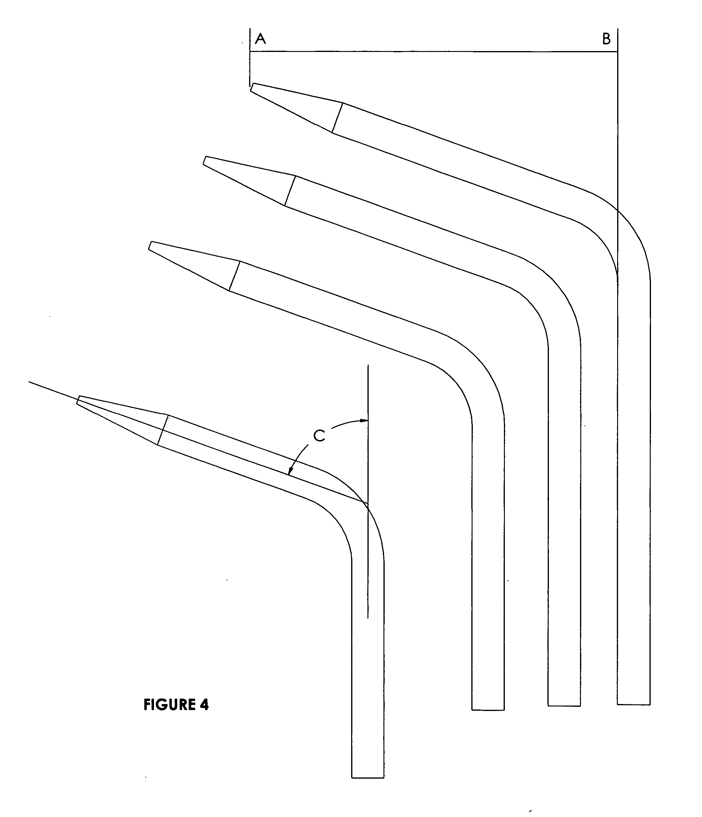
[0138]FIG. 4 is a partial schematic view of four different embodiments of the urethral sound of the present invention, illustrating different “throws” availa...


PUM


Abstract

A catheter capture device comprising a urethral sound and a sleeve that utilizes balloon inflation to capture the catheter. A catheter capture device comprising a urethral sound and a clamshell device. The clamshell device comprises two halves, one of which comprises two pins. One pin passes through the lateral holes in the tip of the catheter, and the other pin fits into a notch in the bottom half of the clamshell device. A catheter capture device comprising a urethral sound, a sleeve and a pin that passes through the lateral holes in the tip of the catheter. A catheter capture device comprising a urethral sound, a wire and a nodule, wherein the nodule captures the catheter by lodging in the tip of the catheter. The nodule could be a ball, hook, crimped wire or similar object. A method of capturing a catheter in an obese or non-obese patient.

Description

CROSS-REFERENCE TO RELATED APPLICATION [0001] This application is a continuation-in-part of U.S. application Ser. No. 10 / 837,879, which was filed on May 3, 2004. The latter application in turn claims the benefit under 35 U.S.C. § 119(e) of U.S. Application No. 60 / 466,959, filed on May 5, 2003. The contents of these applications are hereby incorporated by reference into the present disclosure.BACKGROUND OF THE INVENTION [0002] 1. Field of the Invention [0003] The present invention relates to the field of medical devices, and more particularly, to a device that facilitates suprapubic catheter placement, even in the morbidly obese, in connection with vaginal surgeries for stress urinary incontinence and pelvic prolapse. The catheter capture device of the present invention can also be used for permanent suprapubic catheterizations in those situations in which patients suffer from incurable incontinence or urinary retention. The catheter capture device of the present invention includes b...

Claims


Application Information

Patent Timeline
IPC IPC(8): A61B17/32A61B17/34A61F2/958A61M5/00A61M5/178A61M25/00A61M27/00H01L21/4763
CPCA61B17/3415A61B17/3417A61M25/0074A61M25/0014A61M25/007A61B2017/320052
Inventor HIGH, KENNETH A.
Owner SWAN VALLEY MEDICAL
Learn More