
Differential PCR-RFLP assay for detecting and distinguishing between nonpathogenic PCV-1 and pathogenic PCV-2

a technology of pcv-2 and rflp, which is applied in the field of detecting and distinguishing porcine circovirus (pcv) infections, can solve the problems of complex study, inability of these tests to detect pcv-2 isolates from different geographic regions, and questionable prior methods' usefulness

Inactive Publication Date: 2005-07-07
VIRGINIA TECH INTPROP INC
View PDF7 Cites 41 Cited by
  • Summary
  • Abstract
  • Description
  • Claims
  • Application Information

AI Technical Summary

Problems solved by technology

The inoculated gnotobiotic piglets developed lesions typical of PMWS, but the study was complicated by the detection of porcine parvovirus (PPV) in inoculated piglets.
However, the ability of these tests to detect PCV-2 isolates from different geographic regions is not known.
The data described herein show that PCV-2 isolates from different geographic regions vary enough in their genomic sequences to indicate that the known techniques will not be consistent in each case and, therefore, the usefulness of the prior methods is questionable.
However, the methods of Lin et al. and Jackwood et al. would not work to differentiate PCV isolates because the PCR-RFLP tests were preferentially designed and nucleotide sequence-specific for IBV.

Method used


Image

  • Differential PCR-RFLP assay for detecting and distinguishing between nonpathogenic PCV-1 and pathogenic PCV-2
  • Differential PCR-RFLP assay for detecting and distinguishing between nonpathogenic PCV-1 and pathogenic PCV-2
  • Differential PCR-RFLP assay for detecting and distinguishing between nonpathogenic PCV-1 and pathogenic PCV-2

Examples


example 1

Isolation of DNA from Tissue Samples

[0042] Various tissue samples (liver, spleen, tonsil, lymph nodes, etc.) were collected from pigs with PMWS as confirmed by immunohistochemistry (IHC). The tissues were stored until use at −80 C. The complete PCV-2 genome was amplified, sequenced and characterized from tissue samples of six selected PMWS cases originated from different geographic regions of North America: two cases from Utah, one from Missouri, one from Iowa, one from Illinois, and one from Canada (Table 1, below). These six PMWS cases, along with four more field cases of PMWS (Table 1, below) from Iowa, were also characterized by the PCR-RFLP analyses.

[0043] DNA was extracted from the various tissue samples with a QIAamp DNA Mini kit (Qiagen, Inc., Valencia, Calif.) according to the protocol supplied by the manufacturer. For each DNA extraction, 25 mg of tissue samples were used. The resulting DNA was eluted in DNase, RNase and proteinase-free water (Eppendorf 5 Primer, Inc., B...

example 2

PCR Amplification of the Complete Genome of PCV-2

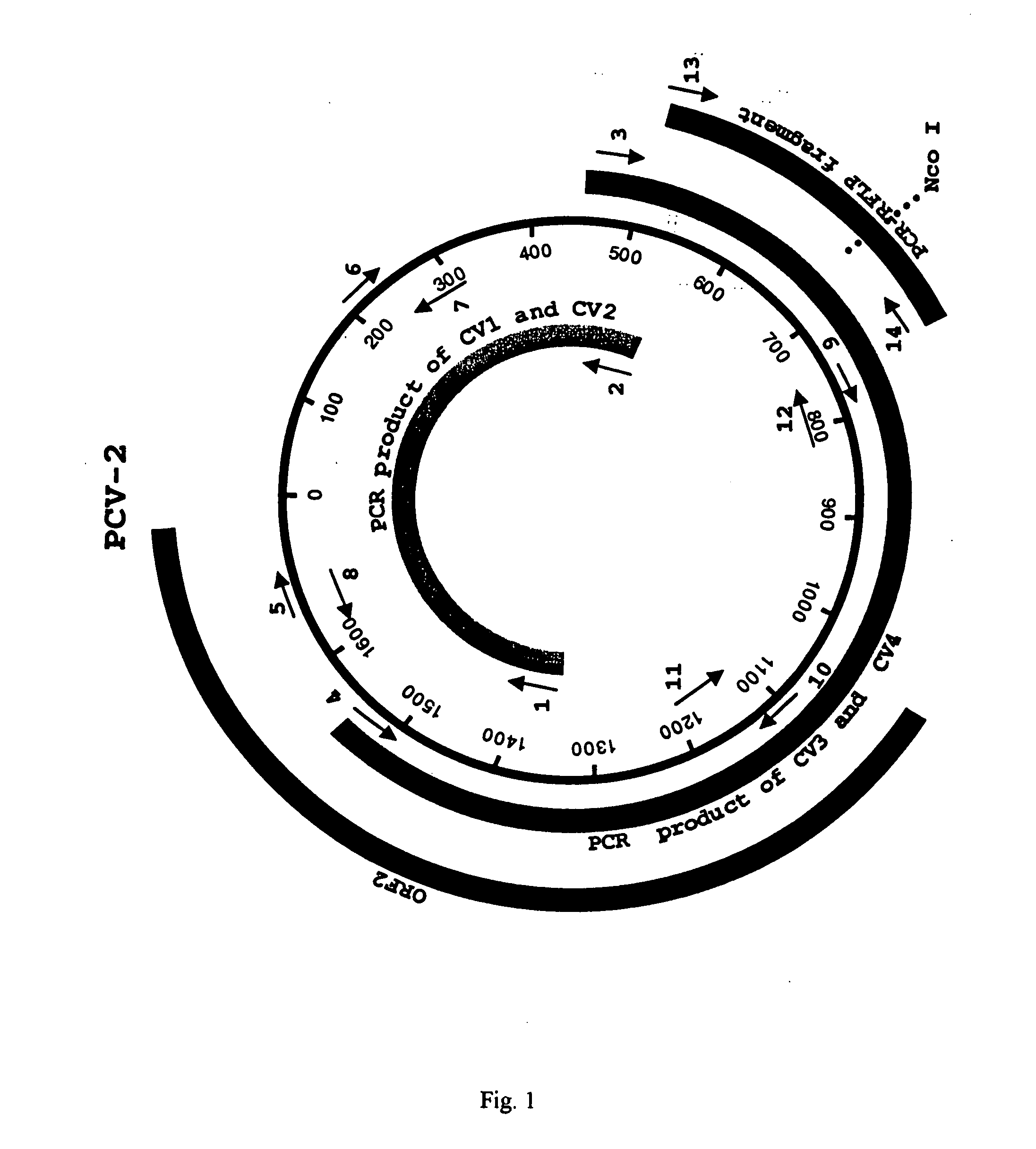
[0044] Two sets of PCR primers were designed on the basis of the published PCV-2 sequence. These primers amplify two overlapping fragments that represent the entire genome of PCV-2 (FIG. 1). The first set of primers, CV1 and CV2 (Table 2, below), amplifies a 989 bp fragment, and the second set of primers, CV3 and CV4 (Table 2, below), amplifies a 1092 bp fragment. The extracted DNA was amplified by PCR using AmpliTaq Gold polymerase (Perkin Elmer, Norwalk, Conn.). The PCR reaction consisted of 35 cycles of denaturation at 94 C for 1 min, annealing at 55 C for 1 min, and extension at 72 C for 3 min, followed by a terminal extension at 72 C for 7 min.

example 3

Nucleotide Sequencing, Sequence and Phylogenetic Analyses

[0045] The PCR products of expected sizes were purified by electrophoresis on a 1% agarose gel followed by extraction with a Geneclean Kit (Bio101, La Jolla, Calif.). Both strands were sequenced with a variety of sequencing primers (Table 2, below) with an ABI automated DNA Sequencer at Virginia Tech's DNA Sequencing Facility. The sequences of the primers used to sequence the complete genome of PCV-2 are listed in Table 2, below and their relative positions in the circular genome are indicated (FIG. 1). The sequences were compiled and analyzed by the MacVector program (commercially available from Oxford Molecular Ltd., Beaverton, Oreg.). The percentages of sequence identity among different PCV isolates were determined with the Clustal alignment program in the MacVector package. Sequence alignments were performed with the ALIGN program in the MacVector package. Phylogenetic analyses were conducted with the aid of the PAUP prog...


PUM

PropertyMeasurementUnit
Lengthaaaaaaaaaa

Abstract

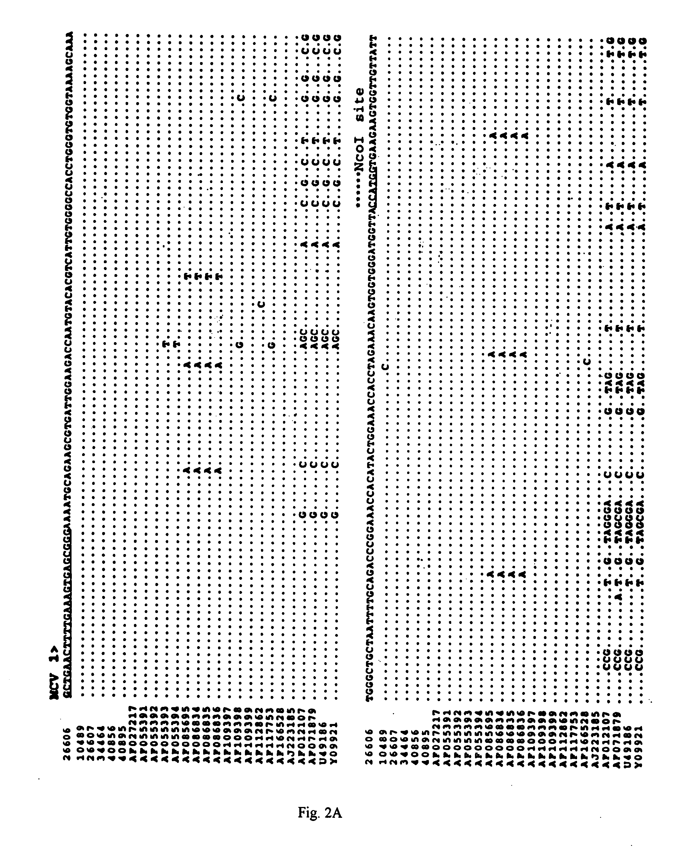
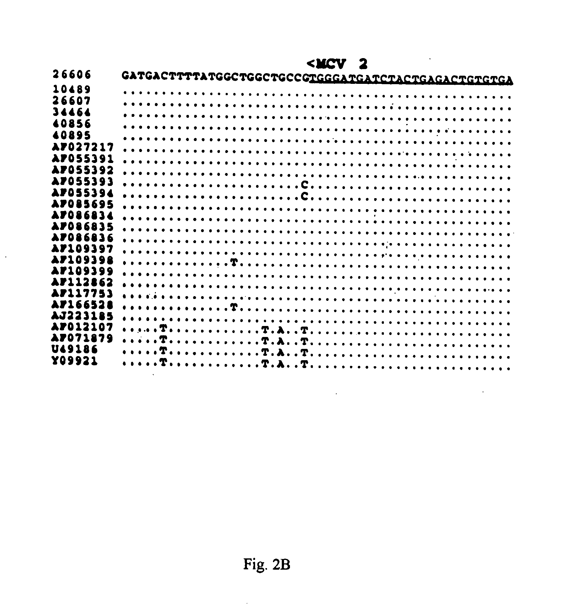
The present invention relates to a method for detecting and differentiating PCV infections in a biological sample taken from a pig which involves amplifying a fragment from an extracted nucleic acid; digesting the fragment with a suitable restriction enzyme such as the unique NcoI restriction enzyme; forming a restriction fragment length polymorphism pattern; and then detecting the presence or absence of a PCV isolate. The invention further concerns the new oligonucleotide primers for differentiating PCV infections comprising a nucleotide sequence selected from the group consisting of MCV1 having a nucleotide sequence set forth in SEQ ID NO:1 and MCV2 having a nucleotide sequence set forth in SEQ ID NO:2. Moreover, this invention provides a novel kit for detecting and distinguishing PCV infections that includes the new oligonucleotide primers and the suitable restriction enzyme.

Description

CROSS-REFERENCE TO RELATED U.S. APPLICATIONS [0001] This application claims the benefit under 35 U.S.C. § 119 (e) of U.S. Provisional Application No. 60 / 301,707, filed Jun. 28, 2001. The prior application is incorporated herein by reference in its entirety.STATEMENT REGARDING FEDERALLY SPONSORED RESEARCH OR DEVELOPMENT [0002] Not Applicable REFERENCE TO A “Sequence Listing”[0003] The material on a single compact disc containing a Sequence Listing file provided in this application is incorporated by reference. The date of creation is ______, 2002 and the size is ______. BACKGROUND OF THE INVENTION [0004] 1. Field of the Invention [0005] The present invention concerns a method for detecting and distinguishing porcine circovirus (PCV) infections by a novel differential polymerase chain reaction-restriction fragment length polymorphism (PCR-RFLP) assay using a restriction enzyme and new primers. The invention further relates to a kit for detecting and distinguishing PCV infections that ...

Claims


Application Information

Patent Timeline
IPC IPC(8): C12Q1/68C12Q1/70
CPCC12Q1/701C12Q1/683
Inventor MENG, XIANG-JINFENAUX, MARTIJN
Owner VIRGINIA TECH INTPROP INC
Learn More