Local magnetic resonance image quality by optimizing imaging frequency

a local magnetic resonance and imaging frequency technology, applied in the field of magnet resonance imaging, can solve the problems of difficult to develop a general shim solution for continually changing heart position and geometry, difficult to achieve uniform field by shim adjustment, image artifacts, etc., to minimize off-resonance related image artifacts, improve image quality, and improve image quality

US20050165295A1Inactive Publication Date: 2005-07-28NORTHWESTERN UNIV
5 Cites 42 Cited by

Patent Information

Authority / Receiving Office
US · United States
Current Assignee / Owner
Publication Date
2005-07-28
Estimated Expiration
Not applicable · inactive patent

Smart Images

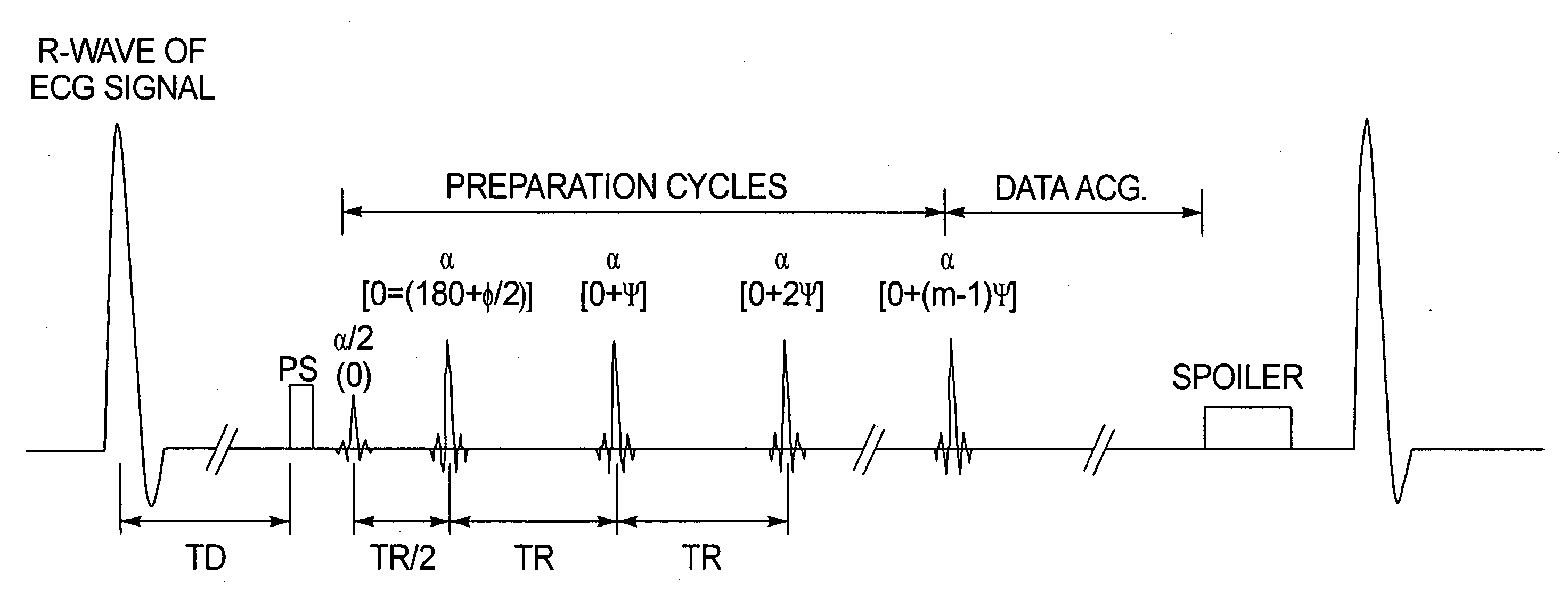
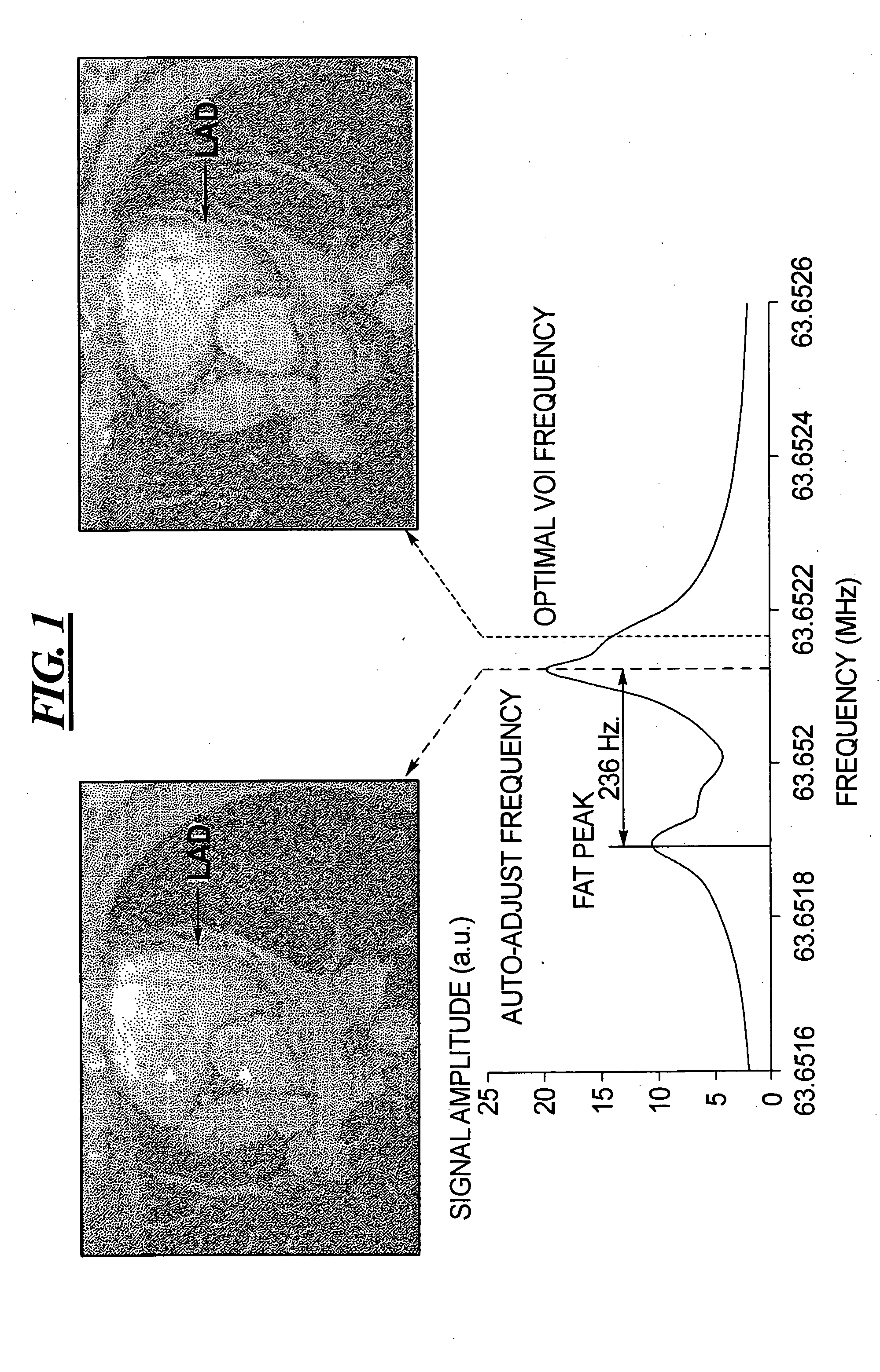
  • Figure 1
    Figure 1
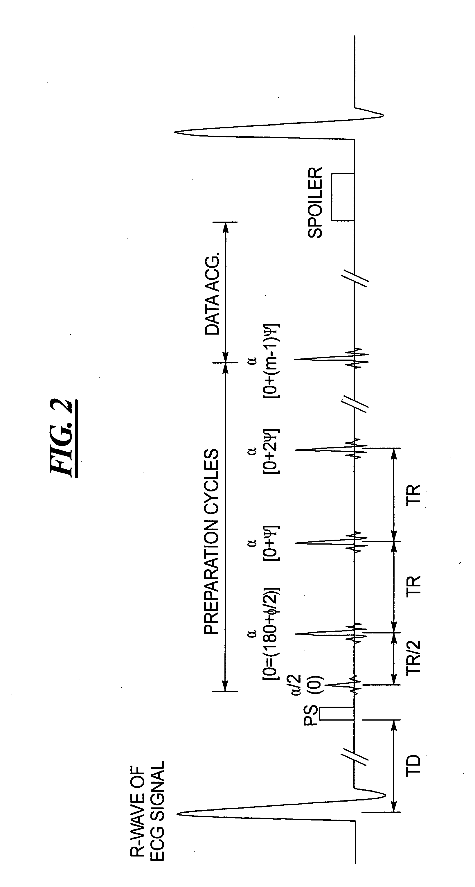
  • Figure 2
    Figure 2
  • Figure 3
    Figure 3
Patent Text Reader

Abstract

In a method and magnetic resonance (MR) imaging apparatus for reducing artifacts due to resonance frequency offsets in a diagnostic MR image, a number of MR scout images of a portion of a subject containing a region of interest (ROI) are generated respectively using different radio frequency (RF) excitation frequencies. Each of the MR scout images has an identifiable image quality in the ROI. The MR scout images are analyzed as to the image quality in the ROI to identify one of the MR scout images having the best image quality in the ROI. An MR diagnostic image is then generated of the portion of the subject containing the ROI, using the RF excitation frequency that was used to generate the MR scout image having the best image quality in the ROI.
Need to check novelty before this filing date? Find Prior Art

Description

STATEMENT REGARDING FEDERALLY SPONSORED RESEARCH

[0001] The United States government has certain rights to this invention pursuant to Grant No. HL-38698 from the National Institutes of Health to Northwestern University.BACKGROUND OF THE INVENTION

[0002] 1. Field of the Invention

[0003] The present invention relates generally to magnetic resonance imaging and, more particularly, to improving local magnetic resonance image quality by optimizing imaging frequency.

[0004] 2. Description of the Prior Art

[0005] With improvements in gradient capabilities in recent years, true fast imaging with steady-state precession (true-FISP) has been successfully used in cardiac cine imaging and coronary artery imaging. In true-FISP, the zeroth moment of the gradients in each TR are zero so that transverse coherences are maintained in successive radio-frequency (RF) cycles. The resonance frequency offset then dominates the phase behavior of the spins and may cause image artifacts. One of the main sour...

Examples

Embodiment Construction

[0024]FIG. 7 schematically illustrates a magnetic resonance imaging (tomography) apparatus for generating a nuclear magnetic image of a subject according to the present invention. The components of the nuclear magnetic resonance tomography apparatus correspond to those of a conventional tomography apparatus, but it is controlled according to the invention. A basic field magnet 1 generates a time-constant, intense magnetic field for polarization (alignment) of the nuclear spins in the examination region of a subject such as, for example, a part of a human body to be examined. The high homogeneity of the basic magnetic field required for the nuclear magnetic resonance measurement is defined in a spherical measurement volume M in which the part of the human body to be examined is introduced. For supporting the homogeneity demands and, in particular, for eliminating time-invariable influences, shim plates of ferromagnetic material are attached at suitable locations. Time-variable influe...