In vitro induction of antigen-specific T-cells using dendritic cell-tumor cell or dendritic cell-viral cell derived immunogens

Inactive Publication Date: 2005-08-04
UNIVERSITY OF PITTSBURGH
View PDF2 Cites 10 Cited by
  • Summary
  • Abstract
  • Description
  • Claims
  • Application Information

AI Technical Summary

Benefits of technology

[0015] There is a need for cancer immunotherapy that provides protective and therapeutic immunity to a wide variety of tumor types. A similar need exists for viral immunotherapy directed towards a wide variety of viral infections.
[0016] The present invention has met the above described needs by providing antigen-specific T-cells and methods of making and using the same. Generally, the Teells are prepared by co-culture of T-ells with formulations comprising dendritic cells and either tumor cells or virally-infected cells. One embodiment of this invention uses a formulation comprising one or more hybridomas; each hybridoma is further comprised of at least one dendritic cell fused to at least one of either a tumor cell or a virally-infected cell. Another embodiment of this invention uses a formulation comprising the products of co-culture of dendritic cells and either tumor cells or virally-infected cells. Both of these formulations produce immunogens specific for the type of tumor or virus used in the formulation. Co-culture of either of these formulations with T-cells results in T-cells that are antigen-specific; the generated T-cells will recognize, and attack, cells expressing the particular antigens with which the T-cells have been co-cultured. In the case of tumor immunotherapy, the generated T-cells provide both protection against tumor challenge and regression of tumor growth. Thus, the T-cells of the present invention provide prophylactic resistance to tumors of the type represented by the tumor cell used in the formulation, and also provide a therapeutic treatment for patients suffering from such tumors. Similarly, in the case of viral immunotherapy, the generated T-cells protect against the viral infection caused by the virally infected cells used in the formulation, and/or provide therapeutic relief for patients having such viral infections.
[0017] Tumor cells and virally infected cells express antigens which can be targeted by T-cells, but the tumor

Problems solved by technology

Tumor cells and virally infected cells express antigens which can be targeted by T-cells, but the tumor cells and virally infected cells themselves do not stimulate T-c

Method used


Image

  • In vitro induction of antigen-specific T-cells using dendritic cell-tumor cell or dendritic cell-viral cell derived immunogens
  • In vitro induction of antigen-specific T-cells using dendritic cell-tumor cell or dendritic cell-viral cell derived immunogens
  • In vitro induction of antigen-specific T-cells using dendritic cell-tumor cell or dendritic cell-viral cell derived immunogens

Examples


example 1

Fusion of DCs and Tumor Cells

[0052] Dendritic cells were prepared from bone marrow as generally described in Celluzzi et al., J. Exp. Med. 183: 283-287 (1996) using GM-CSF as described in the reference. Briefly, bone marrow cells were depleted of lymphocytes and cultured at 5×10⁵ cells / ml in 10% FCS-containing RPMI 1640, obtained from Irvine Scientific, Santa Ana, Calif., with granulocyte macrophage-colony stimulating factor (GM-CSF), in a concentration of 103 U / ml, obtained from Sigma Chemical Company, St. Louis, Mo. Loosely adherent cells were collected on day 6 for fusion. Between about 50 and 75% of the DCs expressed CD86 (B7.2) and Class II MHC (I-A+) antigens, as determined by flow cytometry.

[0053] Day 6 DCs were fused with either B16 or 3LL cells at a ratio of 6:1, DC to tumor cells, using polyethylene glycol at 37° C. After washing by centrifugation, fused cells were cultured overnight at 37° C. in RPMI 1640 (10% FCS).

example 2

Preparation of Dendritic Cell and Tumor Cell Co-cultures

[0054] Dendritic cells were prepared according to the methods of Example 1. Day 6 dendritic cells were then used to form a co-culture with either B16 cells or 3LL cells. Each DC / tumor cell co-culture was prepared by placing DCs into test tubes with the respective tumor cells. A pellet of cells was formed by centrifugation. The pellet was then diluted with RPMI (10% FCS) and incubated overnight at about 37° C. in a 5% CO2 incubator. The ratio of DCs:tumor cells was about 6:1. The products of the co-culture were prepared to further evaluate whether tumor antigens were present in close association with DCs and to evaluate if soluble factors released from the tumors were present on the DCs.

example 3

Efficiency of Fusion and Co-Culture

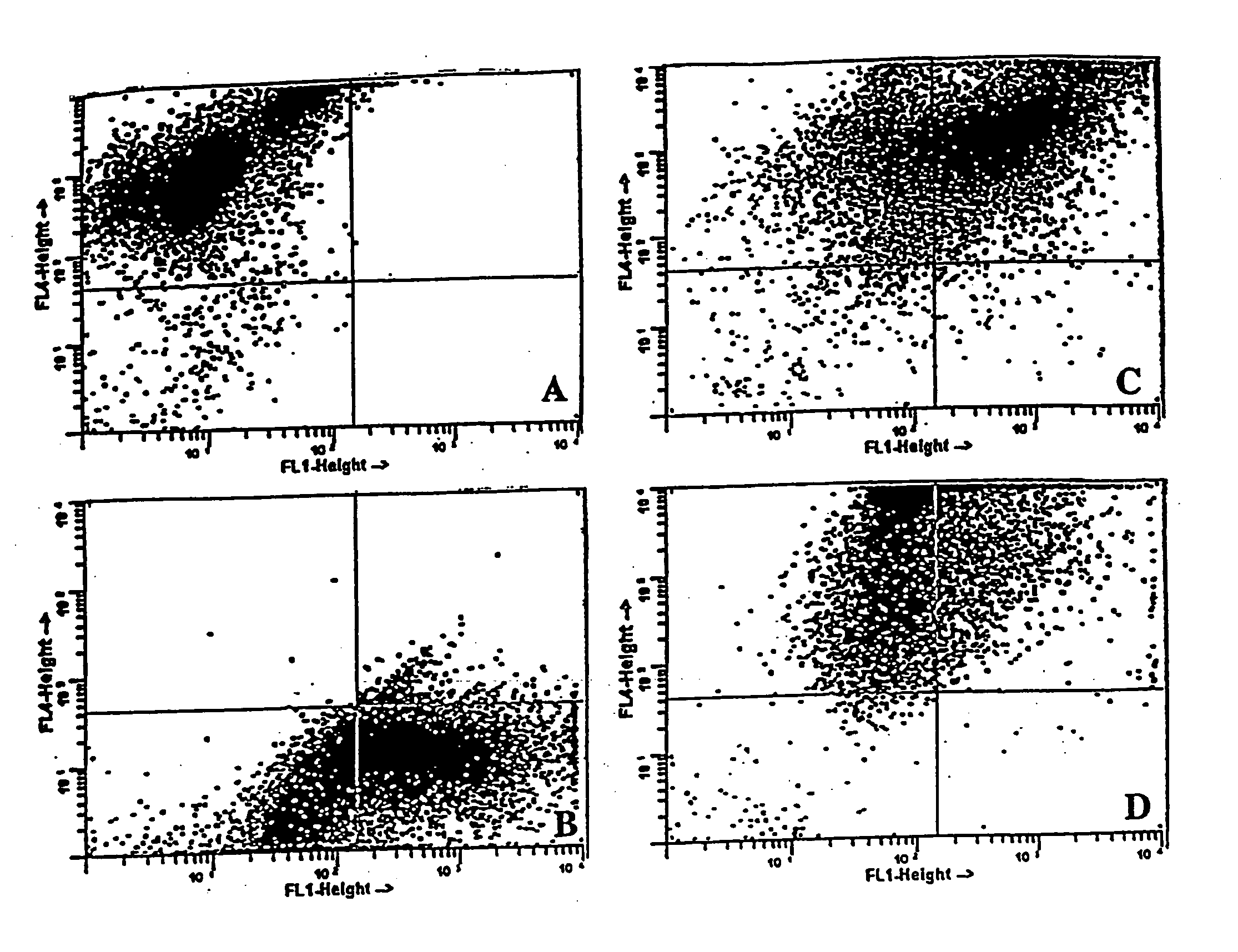
[0055] To determine the efficiency of fusion and co-culture, each of the cell types—DCs, B16 and 3LL—was stained with a different lipophilic fluorochrome before fusion and analyzed using flow cytometry. The tumor cells were stained with DiO while the DCs were stained with Dil, both of which were obtained from Molecular Probes, Inc., Eugene, Oreg. After extensive washing, cells were fused or co-cultured and allowed to incubate overnight at 37° C. Harvested cells were then fixed in 2% paraformaldehyde and the forward and side scatter patterns measured on a Becton Dickinson Facstar Plus with Argon / HeNe duel laser, available from Becton Dickinson Immunocytometry Systems, San Jose, Calif.

[0056] The scatter pattern of each cell type is depicted in FIG. 1. Individual cell staining shows two distinct patterns of DCs (DiI) which shift up (upper left quadrant of FIG. 1A) or B16 tumor cells (DiO) which shift right (lower right quadrant of FIG. 1B) as compar...


PUM

PropertyMeasurementUnit
Timeaaaaaaaaaa
Ratioaaaaaaaaaa

Abstract

Antigen-specific T-cells prepared by culturing T-cells in formulations comprising combinations of DCs and either tumor cells or virally infected cells are disclosed. These formulations generally comprise hybridoma of at least one dendritic cell fused to either at least one tumor cell or at least one virally infected cell, or co-cultures of dendritic cells and either tumor cells or virally infected cells. The resulting T-cells can then be used in immunotherapy methods through adoptive transfer of autologous antigen-specific T-cells into patients using well-established techniques, as agents to identify tumor antigens, and to establish animal models.

Description

CROSS REFERENCE TO RELATED APPLICATIONS [0001] This application is a continuation-in-part of U.S. Ser. No. 09 / 030,985 filed Feb. 26, 1998, which claims priority to U.S. Ser. No. 60 / 039,472, filed Feb. 27, 1997.FIELD OF THE INVENTION [0002] The present invention generally relates to antigen-specific T-cells and methods for making and using the same. More specifically, the present invention relates to antigen-specific T-cells that have been generated by co-culture with immunogens derived from a formulation comprising either hybridomas of dendritic cells and tumor cells or co-culture products of dendritic cells and tumor cells. Alternatively, virally infected cells can be used instead of tumor cells in these hybridomas and co-cultures. The use of these T-cells as prophylactic and therapeutic agents against tumors and viral infection is also the subject of the present invention. BACKGROUND OF THE INVENTION [0003] T-cells, including cytotoxic T-lymphocytes (CTLs), are a critical componen...

Claims


Application Information

Patent Timeline
IPC IPC(8): A61K39/00C12N5/0783C12N5/16
CPCA61K39/0011A61K2039/5154A61K2039/5156A61K2039/5158C12N2502/99C12N5/16C12N2501/22C12N2501/23C12N2502/30C12N5/0636
Inventor FALO, LOUIS D. JR.STORKUS, WALTER
Owner UNIVERSITY OF PITTSBURGH
Why Eureka
Try Eureka