Method for inhibiting tumor formation and growth
a tumor and growth technology, applied in the field of tumor formation and growth inhibition, can solve the problems of tightly controlled angiogenesis and limited application prospects
- Summary
- Abstract
- Description
- Claims
- Application Information
AI Technical Summary
Problems solved by technology
Method used
Image
Examples
example 1
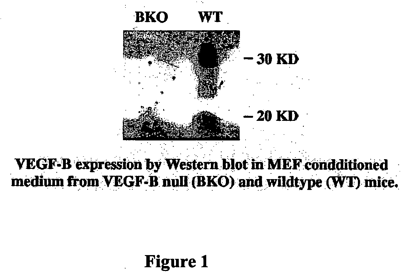
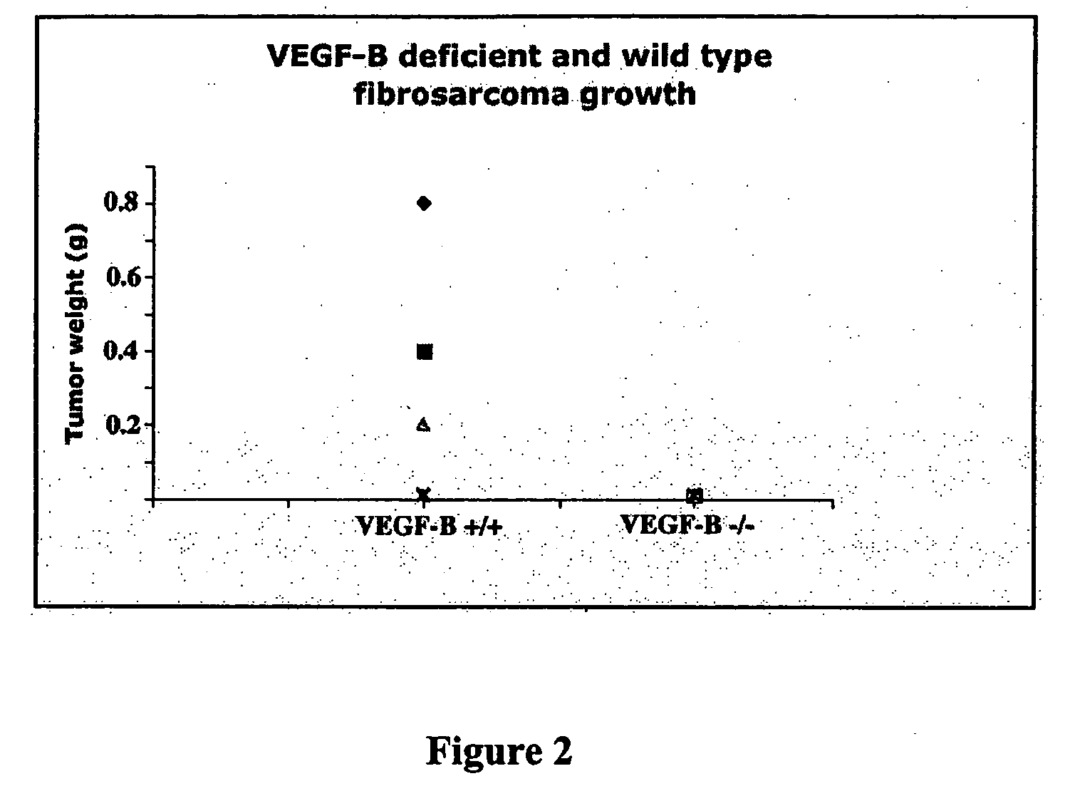
In Vitro Growth and Transformation of MEFs Lead to Induction of VEGF-B Expression, and VEGF-B is Required to Initiate and Support Tumor Growth
[0080] 1. Cell Lines and Cell Culture
[0081] Mouse embryonic fibroblast cells (MEFs) from VEGF-B deficient (1) and wild type embryos (E12) were isolated as described previously (2). The cells were grown in DMEM medium (Life Technologies, Inc.) with 10% fetal bovine serum (Life Technologies, Inc.), glutamine, and penicillin / streptomycin. The MEFs were immortalized after co-transfection with SV40 T antigen (3), and a hygromycin resistance gene, and selected in hygromycin-containing medium (120 μg / ml) for one week. The resistant MEFs were then infected with H-ras61L retrovirus, and selected the next day in puromycin-containing medium (4 μg / ml) for one week (The plasmids and retrovirus were kindly provided by Dr. P. Carmeliet of the Flanders Interuniversity Institute for Biotechnology).
[0082] All transformed cells, including the MEF cells from V...
example 2
Tumor Growth is Normal in VEGF-B-Deficient Mice and Up-Regulation of VEGF-B Stimulates Tumor Growth
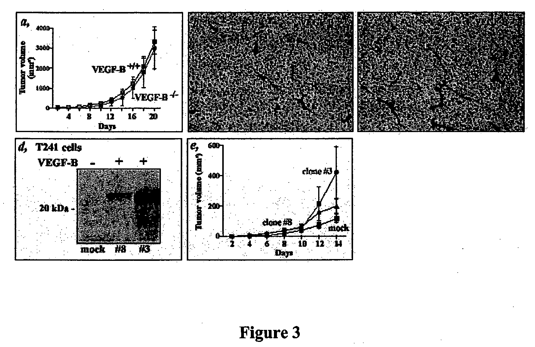
[0092] Murine T241 fibrosarcoma cells (which naturally express both the 167 and 186 isoforms of VEGF-B) were transplanted (1×10⁶ cells) into both wild-type (+ / +) and VEGF-B KO mice (− / −) and the growth of the tumor was followed. As shown in FIG. 3(a), there was no difference is tumor growth, indicating that the host VEGF-B does not affect tumor growth.
[0093] An antibody to PECAM was used to stain tissue sections from the transplanted tumors. As shown in FIGS. 3(b) and (c) there was no difference in vessel density in cells transplanted into either WT (for VEGF-B) or VEGF-B KO mice.
[0094] Murine VEGF-B167 was cloned into the pcDNA3.1 expression vector which was transfected into T231 cells using lipofectin. Clones were selected using neomycin (400 micrograms / ml). FIG. 3(d) shows a western blot using an antibody to VEGF-B (which recognizes both 186 and 167 isoforms (see Aase et al., 199...
example 3
Monoclonal Antibodies Against VEGF-B Have Antagonistic Effects and Prevent Binding of VEGF-B to VEGFR-1
[0096] A. The Specificity of the Anti-VEGF-B Monoclonal Antibody MAB3372
[0097] Microtiter plates (Nunc Maxisorp) were coated with 1.0 μg / ml of recombinant VEGF-B protein (from Amrad Corp., Melbourne, Australia) in 50 μl 100 mM NaHCO3 for overnight at 4° C. Residual binding capacity of the plates were blocked by incubating with PBS, supplemented with 3% BSA, for 0.5 hr at room temperature. The monoclonal anti-VEGF-B antibody MAB3372 (R&D Systems, Minneapolis) was diluted in 1% BSA in PBS at various concentrations, ranging from 0.03-1.0 μg / ml, and allowed to bind for 2 hr. Bound antibodies were incubated with 50 μl anti-mouse-IgG, conjugated with alkaline phosphatase (Sigma-Aldrich, 1:3000) for 2 hr. For visualization of the enzyme-conjugated secondary antibodies, 75 μl of NPP substrate (Sigma-Aldrich) was added. After stopping the reaction with 25 μl 0.5 M NaOH, the absorbance was...
PUM
| Property | Measurement | Unit |
|---|---|---|
| temperature | aaaaa | aaaaa |
| concentrations | aaaaa | aaaaa |
| molecular mass | aaaaa | aaaaa |
Abstract
Description
Claims
Application Information
 - R&D
- Intellectual Property
- Life Sciences
- Materials
- Tech Scout
- Unparalleled Data Quality
- Higher Quality Content
- 60% Fewer Hallucinations
Browse by: Latest US Patents, China's latest patents, Technical Efficacy Thesaurus, Application Domain, Technology Topic, Popular Technical Reports.
© 2025 PatSnap. All rights reserved.Legal|Privacy policy|Modern Slavery Act Transparency Statement|Sitemap|About US| Contact US: help@patsnap.com


