Medical image processing apparatus and medical image processing method

a technology of medical image and processing apparatus, applied in the field of medical image processing apparatus and medical image processing method, can solve the problems of unstable identification of a region of interest, insufficient search, long search time, etc., and achieve the effect of high accuracy

Inactive Publication Date: 2005-10-27
PANASONIC CORP
View PDF4 Cites 44 Cited by
  • Summary
  • Abstract
  • Description
  • Claims
  • Application Information

AI Technical Summary

Benefits of technology

[0007] The present invention has been conceived in view of the above problems, and it is an object of the present invention to provide a medical image process...

Problems solved by technology

This causes a problem that the accuracy of processing such as the identification of a region of interest becomes unstable, since the similarity can be low between the dictionary image and an ultrasound image being compared.
Furthermore, while the above conventional arts are capable of narrowing the search range with reference to the position of a valve, it is possible that a search is not performed appropriately or that it takes a long time for maki...

Method used


Image

  • Medical image processing apparatus and medical image processing method
  • Medical image processing apparatus and medical image processing method
  • Medical image processing apparatus and medical image processing method

Examples


first embodiment

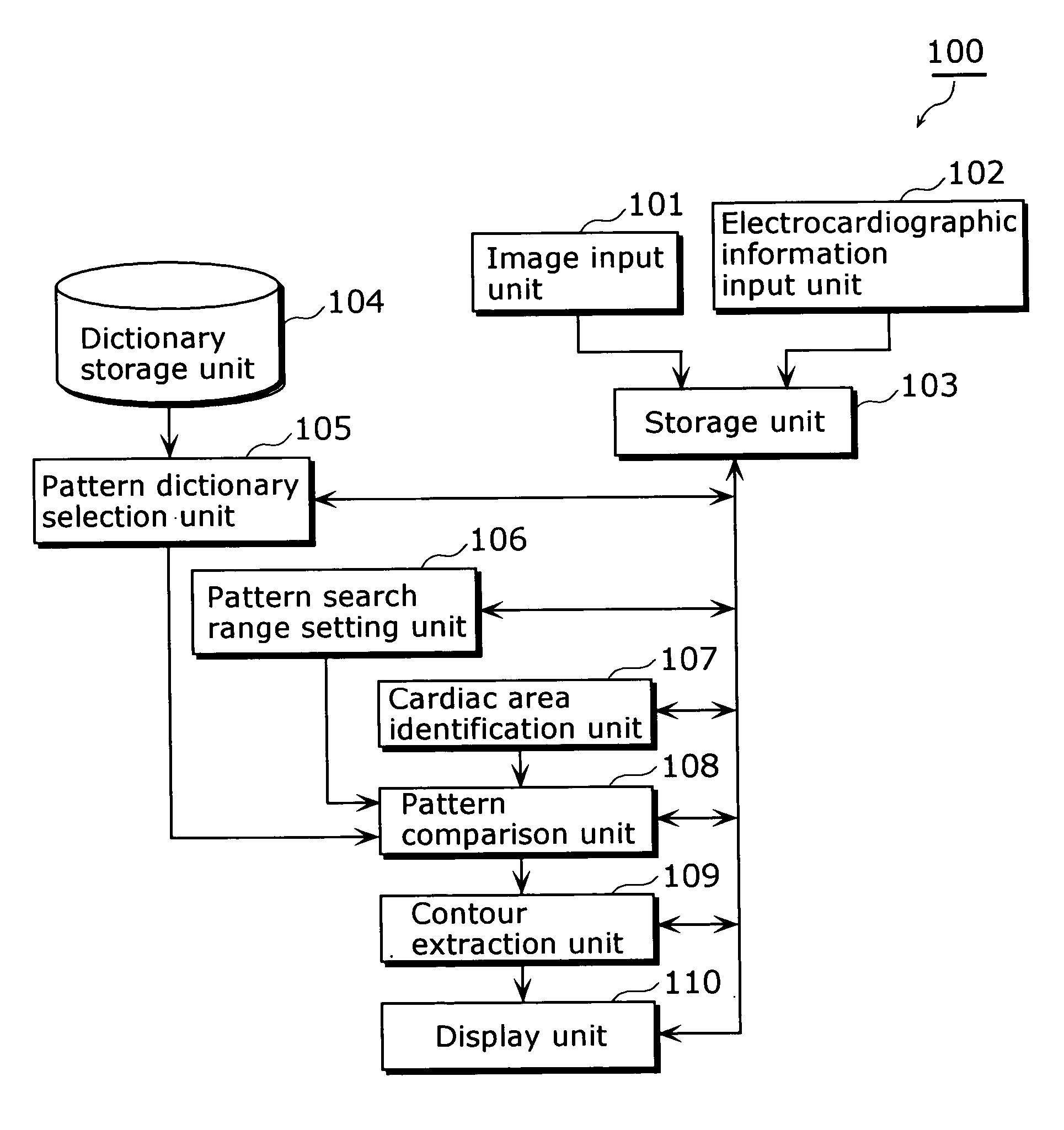
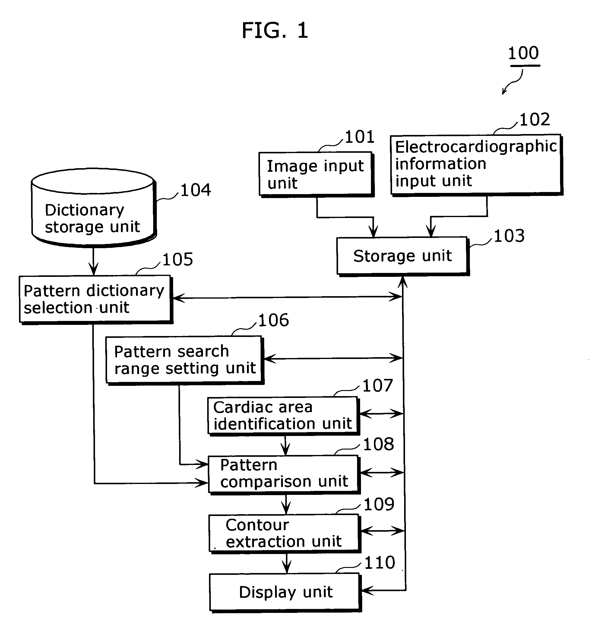
[0029]FIG. 1 is a block diagram showing a functional structure of a medical image processing apparatus 100 according to the first embodiment. The medical image processing apparatus 100 is a diagnostic apparatus for medical use, such as an ultrasonic diagnostic apparatus that generates ultrasound images based on echo signals of ultrasonic pulses emitted to a subject, an X-ray CT apparatus that generates tomograms based on the amount of X rays passing through a subject, and an MRI apparatus that generates magnetic resonance (MR) images based on electromagnetic waves released from a subject. The medical image processing apparatus 100 extracts an internal contour of an organ (e.g. heart and blood vessel) whose activity is cyclic, using moving images of such organ and an electrocardiogram (ECG) obtained from the subject, and displays the extracted contour or the like. Note that, for convenience sake, the following descriptions are provided on the assumption that the medical image process...

second embodiment

[0045] The first embodiment describes an embodiment in which a pattern comparison is performed using a previously prepared dictionary image. The second embodiment describes an embodiment in which pattern comparison is performed using an ultrasound image or the like that is obtained in real-time at examination time.

[0046]FIG. 8 is a block diagram showing a functional structure of a medical image processing apparatus 200 according to the second embodiment. The medical image processing apparatus 200, an example of which is an ultrasonic diagnostic apparatus as in the case of the medical image processing apparatus 100 of the first embodiment, is an imaging diagnostic apparatus that extracts an inner contour of a heart or the like using an ECG and moving images of the heart generated at the speed of 30 frames per second, and displays the extracted contour or the like. Note that the components that are the same as those described in the first embodiment are assigned the same numbers and ...

third embodiment

[0058]FIG. 11 is a block diagram showing a functional structure of a medical image processing apparatus 300 according to the third embodiment. The third embodiment describes an example usage of a contour extraction method, and the present medical image processing apparatus 300 is an imaging diagnostic apparatus that accepts ultrasound images of a heart or the like and extracts and displays an inner contour of the heart.

[0059] Referring to FIG. 11, the input unit 101 accepts image data. The storage unit 103 holds the image data. The characteristic position specification unit 201 accepts an operator's specification of the position of each characteristic area. The contour extraction unit 109 extracts an inner contour of a ventricle based on the specified characteristic positions and images stored in the storage unit 103. The display unit 110 displays the extracted contour, the image information and the like.

[0060] Next, a description is given of operations of the medical image proces...


PUM


Abstract

In a medical image processing apparatus 100 that is capable of extracting a contour of an organ or the like in a highly accurate manner within a short time, an image input unit 101 accepts ultrasound images or the like. An electrocardiographic information input unit 102 accepts electrocardiographic information (e.g. ECG waveform). A storage unit 103 stores image data representing the ultrasound images and the electrocardiographic information in association with each other. A dictionary storage unit 104 stores image patterns (dictionary images) used for pattern matching. A pattern dictionary selection unit 105 selects a dictionary image used for pattern matching with reference to the electrocardiographic information. A pattern search range setting unit 106 specifies a range in which pattern matching is to be performed, based on the electrocardiographic information. A cardiac area identification unit 107 detects a cardiac area in the image. A pattern comparison unit 108 identifies the position of each characteristic area in the image by pattern matching. A contour extraction unit 109 extracts a contour based on the position of each characteristic area and the ultrasound image. A display unit 110 displays the extracted contour.

Description

BACKGROUND OF THE INVENTION [0001] (1) Field of the Invention [0002] The present invention relates to a medical image processing technology, and particularly to an image processing technology for identifying a region of interest, extracting a contour, and the like from medical images of an organ or the like such as a heart whose activity is cyclic, the medical images being obtained on a time series basis. [0003] (2) Description of the Related Art [0004] Conventionally, in the case where a region of interest is identified in and the contour of an organ or the like is extracted from an image of a living body (e.g. ultrasound image and X-ray computed tomogram (CT) image), the spatial position of the obtained ultrasound image is identified first by means of pattern matching or the like. Then, pattern matching and a specification of each characteristic area using a pointing device are performed in combination, in addition to binarization, edge detection, or the like. The above pattern ma...

Claims


Application Information

Patent Timeline
IPC IPC(8): G01R33/28A61B5/00A61B5/04A61B5/055A61B5/107A61B6/03A61B8/00A61B8/06A61B8/08A61B8/12A61B8/14G01R33/54G06K9/00G06T1/00G06T5/00G06T7/20G06T7/60
CPCA61B5/1075A61B6/503A61B6/504A61B8/06A61B8/0891A61B8/0883G06T7/0083G06T7/2006G06T2207/10132G06T2207/30048G06T7/0012G06T7/12G06T7/215
Inventor YODEN, SADATO
Owner PANASONIC CORP
Why Eureka
Try Eureka