

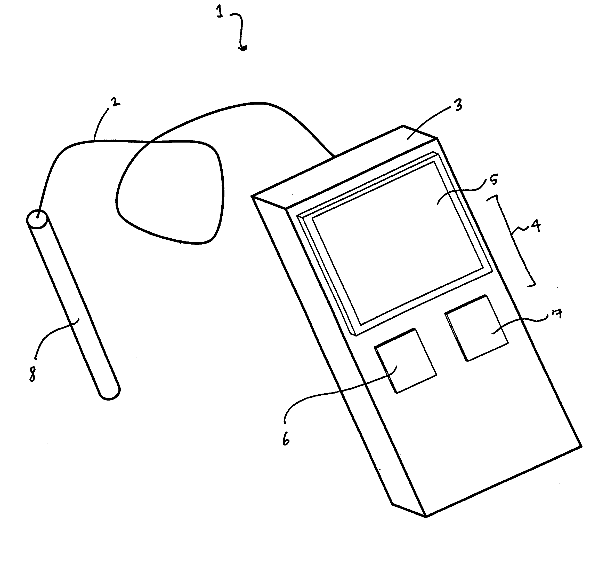
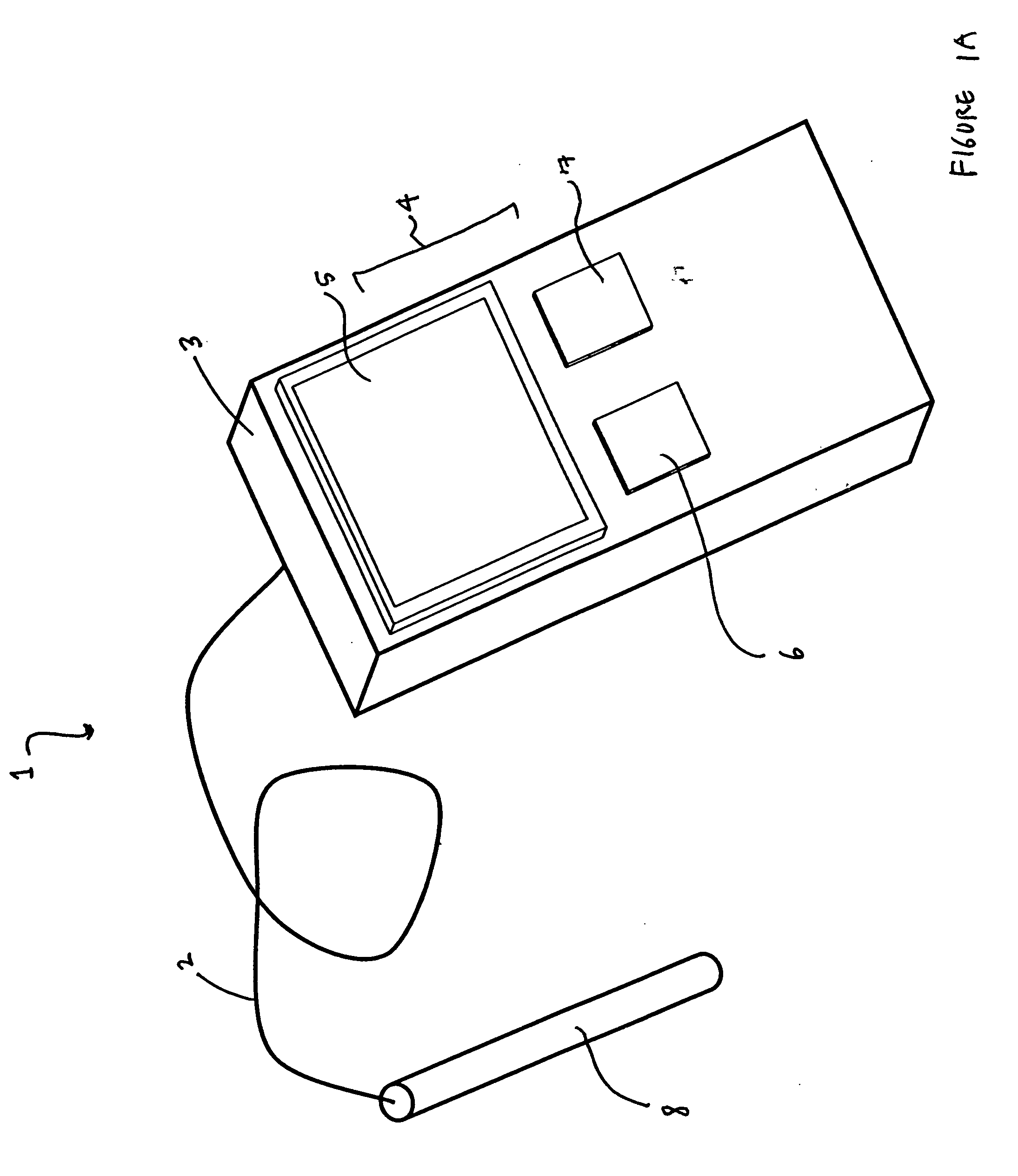
Insertable ultrasound probes, systems, and methods for thermal therapy
a technology of insertable ultrasound and probes, applied in the field of medicine, can solve the problems of insufficient targeting of energy delivery and often sustained trauma, and achieve the effect of raising the temperature of native tissu
- Summary
- Abstract
- Description
- Claims
- Application Information
AI Technical Summary
Benefits of technology
Problems solved by technology
Method used
Image

Examples
embodiment 1
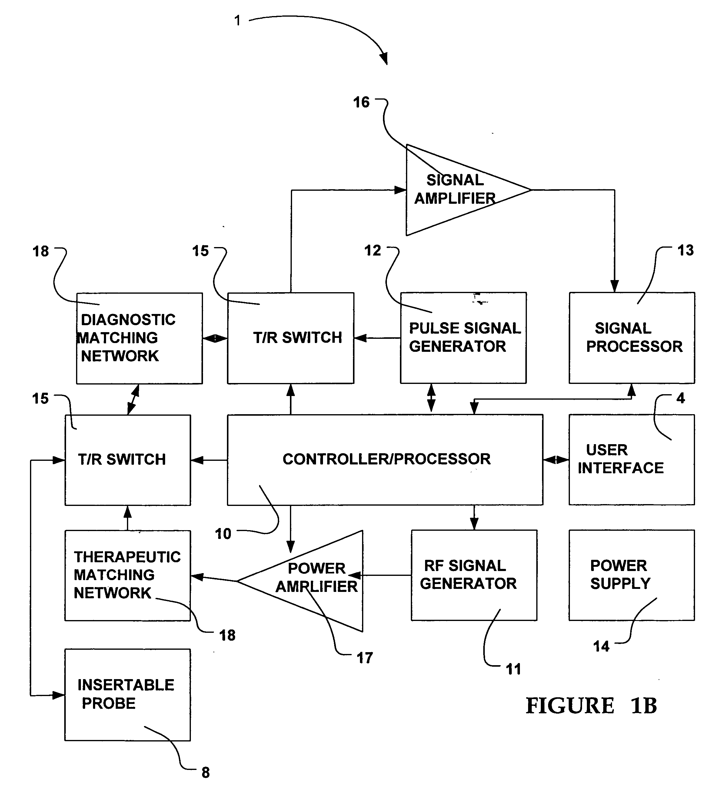
[0036]FIGS. 2A, 2B and 2C illustrate one embodiment of insertable probe 8. As best illustrated in FIG. 2A, insertable probe 8 is comprised of an elongated shaft 20. Elongated shaft 20 is comprised of proximal section 21, distal section 22, and distal tip 23, located at the distal extremity of distal section 22. Elongated shaft 20 is generally a hollow tube comprising a lumen 24 and is generally adapted to house ultrasound transducer assembly 25, as well as any and all other various electronic connectors, cables, and / or wires (collectively illustrated by reference no. 26) needed to operationally interconnect transducer assembly 25 to the rest of therapeutic system 1 and device 3. Alternatively, shaft 20 may be comprised of a solid materials and the various other component being embedded in said material.
[0037] Elongated shaft 20 may be fabricated from any thermally conductive material having high specific heat characteristics (such as copper, brass, nitinol, or other like materials)...
embodiment 2
[0043]FIGS. 3A-3B illustrate another embodiment of probe 8 wherein a “wave guide” design is used. In this embodiment, the emitted acoustic beams from transducer assembly 25, located at proximal section 21, are transmitted, or guided, down elongated shaft 20. The acoustic waves (depicted by arrows), generated by transducer assembly 25, are propagated down elongated shaft 12 and emitted from the distal tip 23 to the tissues.
[0044] As shown, transducer assembly 25 is coupled to elongated shaft 20 via a stand-off means 30 outside of, or external to, elongated shaft 20. As will be appreciated by those skilled in the art, stand-off means 30 and elongated shaft 20 should be fabricated from materials with low attenuation characteristics in order to allow for the efficient transmission of acoustic waves into, and down, elongated shaft 20. However, the acoustic velocity of these materials should differ, thus allowing the emitted acoustic waves (depicted by the arrows) to be bent, or refracte...
embodiment 3
[0046] Referring to FIGS. 4 and 5, an additional embodiment of insertable probe 8 is provided. In this embodiment, probe 8 is adapted for use in conjunction with guidewire 50 (a device typically used in various percutaneous coronary, peripheral, and neurovascular transcatheter procedures). In this embodiment, probe 8 is comprised of one or more lumens. In FIG. 5, a dual lumen configuration is shown wherein inner lumen 51 and outer lumen 52 extend longitudinally from distal tip 23 of elongated shaft 20 to the proximal end or section 21 of elongated shaft 20. Inner lumen 51 is provided so that guidewire 50 (or other like device) may be threaded through it and used to advance probe 8 to an operative location inside the patient. Inner lumen 51 is comprised of a first exit port (not shown) positioned at proximal section 21 of elongated shaft 20 and a second exit port 53, preferably positioned in distal shaft section 22. These ports allow for the guidewire to be threaded through and out p...
PUM
 Abstract
Description
Claims
Application Information