

Microneedle devices and microneedle delivery apparatus
a technology of microneedle and microneedle, which is applied in the direction of infusion needles, other medical devices, surgery, etc., can solve the problems of compromising the ability of microneedle devices, affecting the effectiveness of microneedle delivery, and affecting the structural integrity of the microneedle itself, so as to enhance the effective perforation of the stratum corneum, reduce the effect of tip fractures and effective perforation
- Summary
- Abstract
- Description
- Claims
- Application Information
AI Technical Summary
Benefits of technology
Problems solved by technology
Method used
Image

Examples
example 1
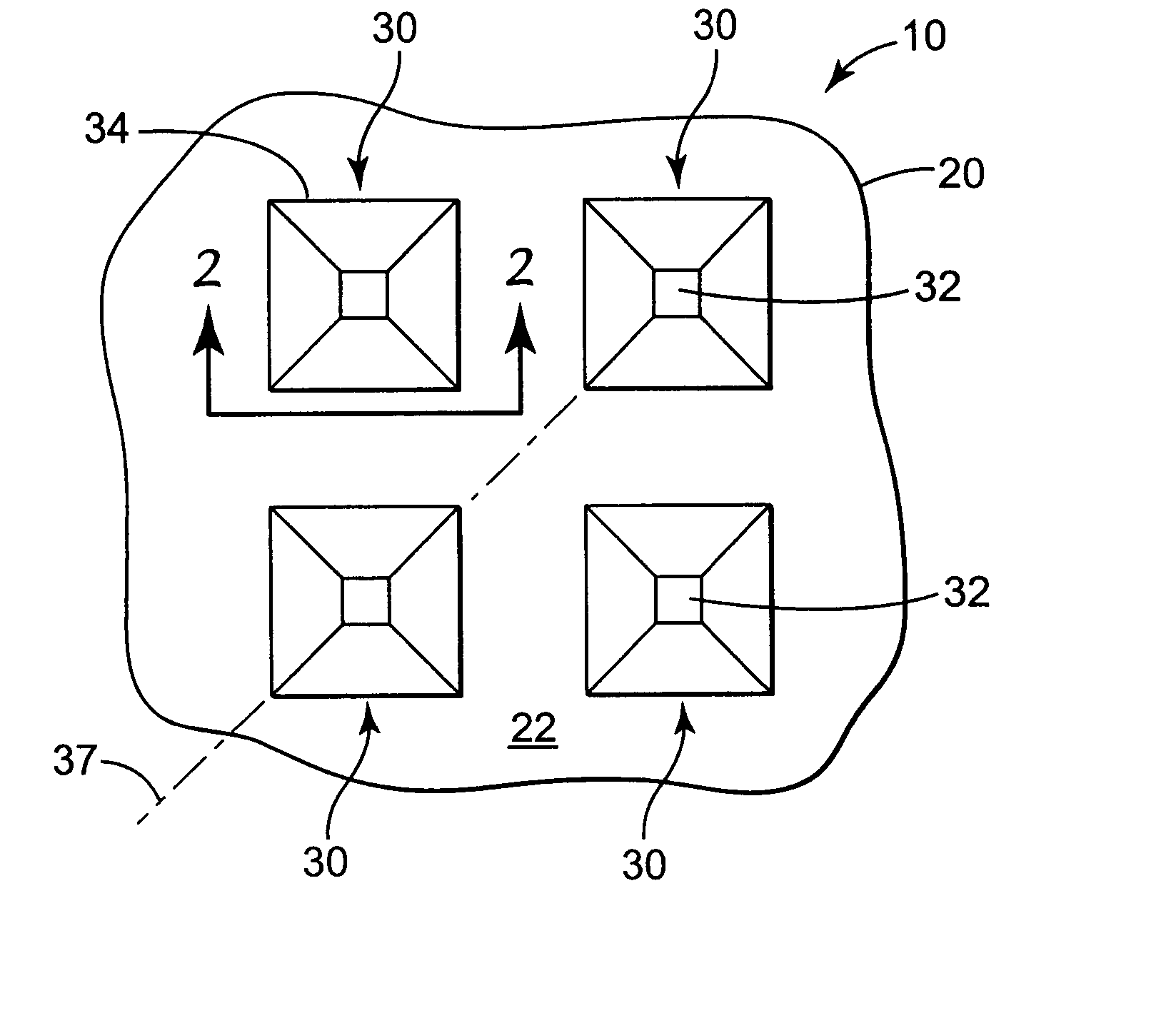
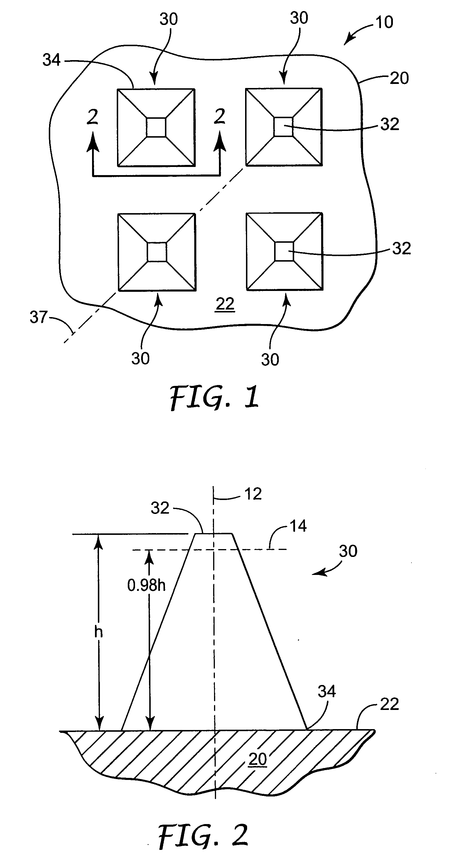
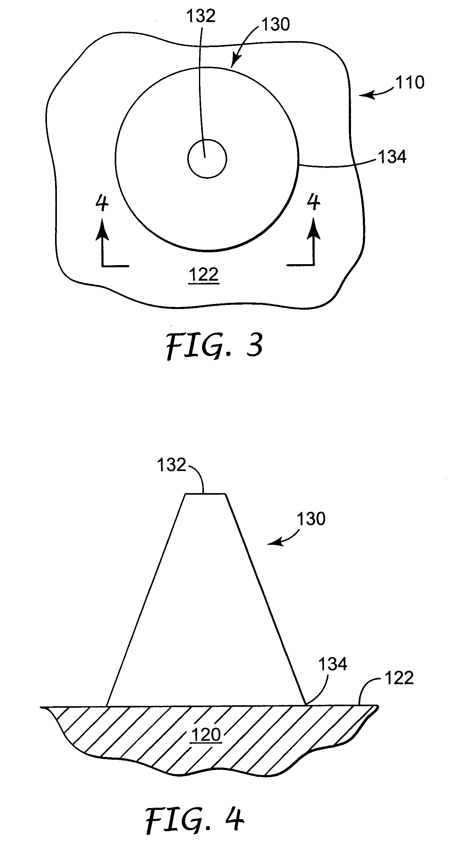
[0095] Microneedle arrays were prepared using the general methods described in U.S. patent application Ser. No. 09 / 947,195, filed on Sep. 5, 2001 and entitled MICRONEEDLE ARRAYS AND METHODS OF MANUFACTURING THE SAME. Two layers of polyimide (KAPTON H, DuPont, Wilmington, Del.) were laminated together to form a mold substrate with a thickness of 250 μm. The mold substrate was laser ablated to form a structured surface with cavities in the shape of the microneedles described below. The structured surface was treated with a seed layer by vapor coating of silver and subsequently electroformed with nickel to form a metallic microneedle array. The thickness of the nickel backplate was approximately 230 μm. The array was removed from the mold and stored prior to use.
[0096] The microneedle arrays were elliptical in shape at the base and tapered to a blunt tip with a surface area of approximately 20 μm 2. The major axis of the base was approximately 100 μm in length and the minor axis of th...
example 2
[0100] Following the method of Example 1, a MINITRAN transdermal patch containing 0.0135 gm nitroglycerin (3M, Maplewood, Minn.) was applied to human cadaver skin treated with the microneedles of Example 1. The human cadaver skin was soaked in 0.025M phosphate buffered saline (PBS) and the receptor solution used was also PBS. Five replicates were performed and average cumulative flux is reported in Table 2. A control sample consisting of the transdermal patch applied to untreated cadaver skin was also tested.
TABLE 2Nitroglycerin Cumulative Flux [μg / cm2]Time [hrs]0246244872Treated0125286447189935944695Untreated060.7197338179835074670
example 3
[0101] Following the method of Example 2, a solution of 0.174 gm / mL sodium ceftriaxone (ROCEPHIN, Roche Labs, New Jersey) in water was applied to human cadaver skin treated with the microneedles of Example 1. Average cumulative flux was determined at 2 hours after application of the solution. At subsequent times, the donor solution began decomposing, as indicated by an increasing red color, so no further time points were sampled. The average cumulative flux at 2 hours for the solution applied to treated skin was 46.6 μg / cm2. The average cumulative flux at 2 hours for the solution applied to untreated skin was 0 μg / cm2.
PUM
 Abstract
Description
Claims
Application Information