

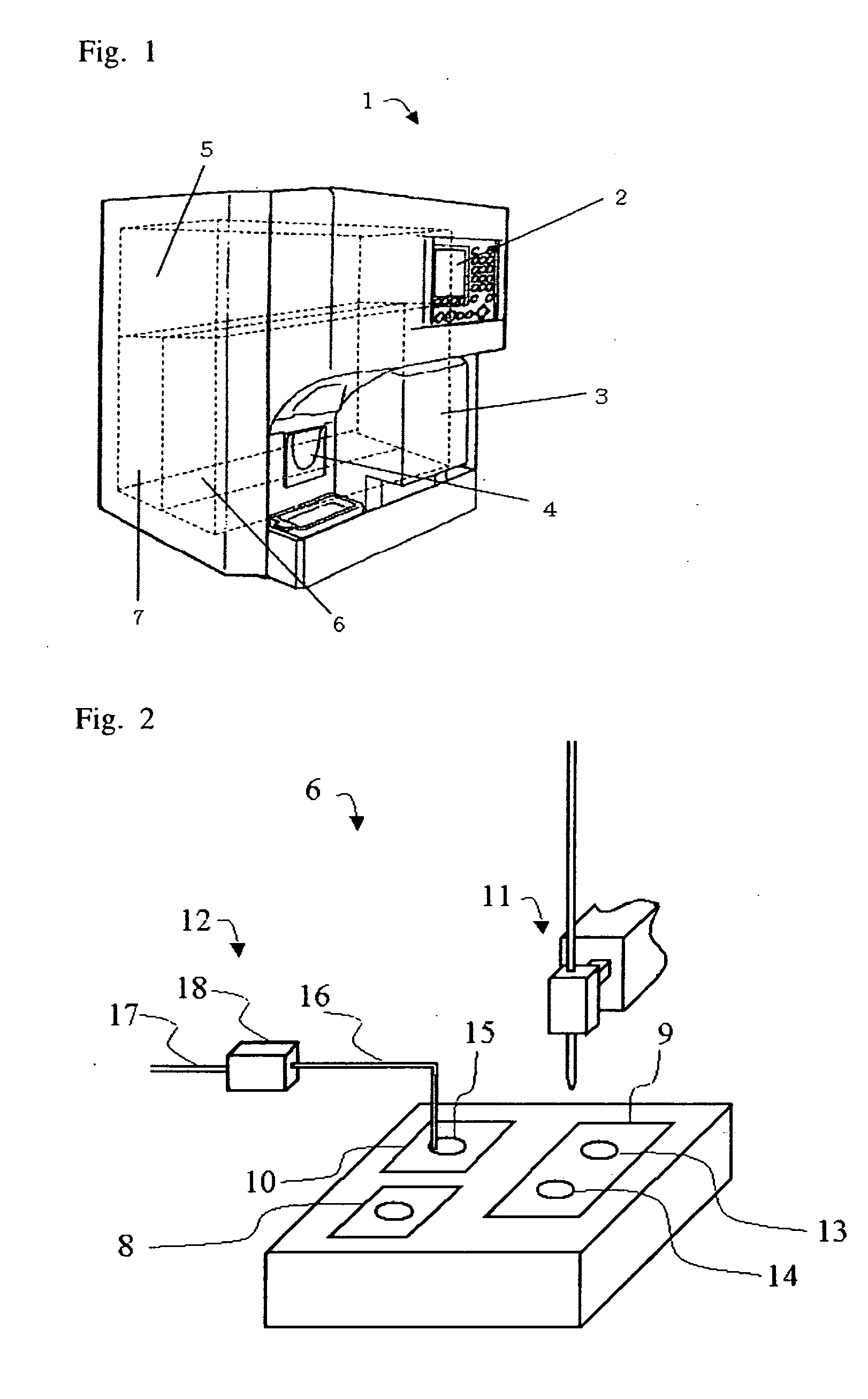
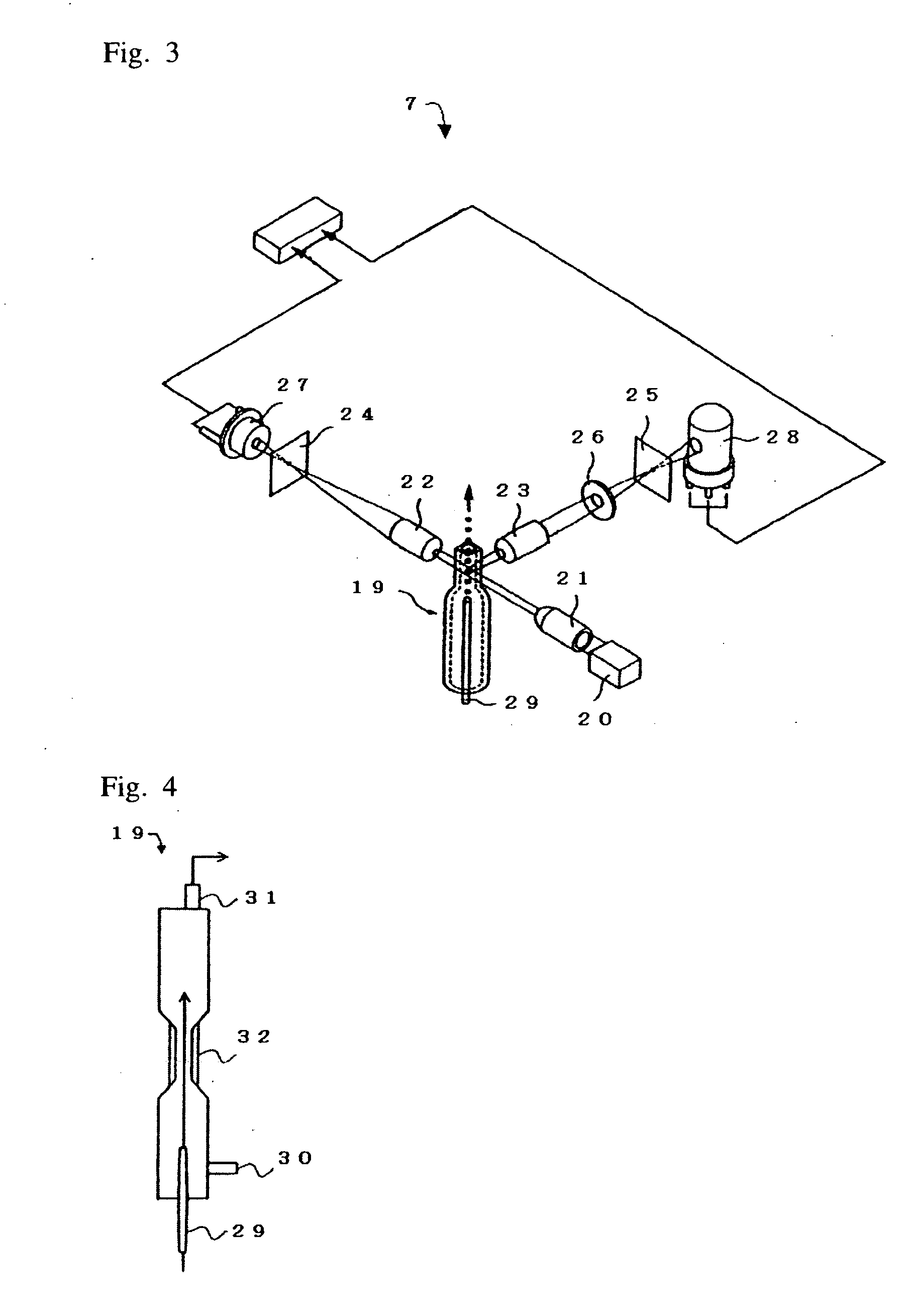
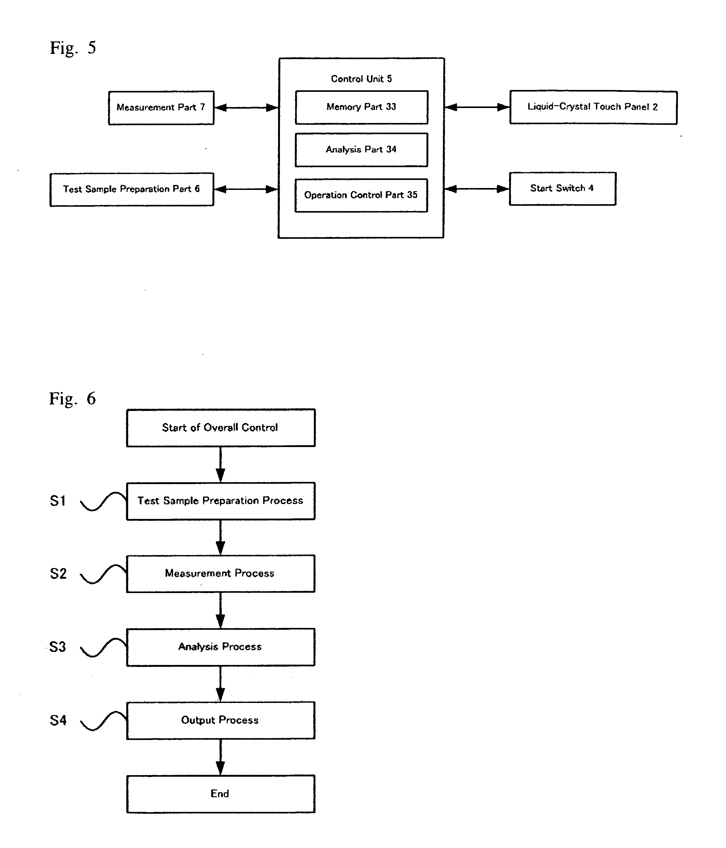
Apparatus and method for cell analysis
a mononuclear cell and apparatus technology, applied in the field of apparatus and method for measuring mononuclear cells, can solve the problems of not revealing the identification of living mononuclear cells including stem cells in the publication, damaged cells necessary for therapy, etc., and achieves the effect of identifying living mononuclear cells in analytes more easily and quickly
- Summary
- Abstract
- Description
- Claims
- Application Information
AI Technical Summary
Benefits of technology
Problems solved by technology
Method used
Image

Examples
example 1
MEASUREMENT EXAMPLE 1
[0052] Two types of human peripheral blood ((I) and (II)) were used as analytes in this measurement example. The different types of peripheral blood were collected from patients with different diseases. The leukocytes of peripheral blood (I) had a very high content of lymphocytes. The leukocytes of peripheral blood (II) had a very high content of granulocytes.
[0053] While collected peripheral blood is allowed to stand at room temperature, the content of dead cells in the peripheral blood generally increases with time. In this measurement example, peripheral bloods (I) and (II) were each collected and allowed to stand at room temperature for 2 or 24 hours before used as analytes.
[0054] The scattergrams as shown in FIGS. 9A, 9B, 10A, and 10B were obtained using the cell analysis apparatus 1 as described above. FIGS. 9A and 9B show the results of the measurement using peripheral blood (I) as an analyte, wherein FIG. 9A shows the result of the measurement in which...
example 2
MEASUREMENT EXAMPLE 2
[0062] In this measurement example, peripheral blood was collected from a healthy subject and allowed to stand at room temperature for 2 or 32 hours before used as an analyte.
[0063] The scattergrams as shown in FIGS. 11A and 11B were obtained using the cell analysis apparatus 1 as described above. FIG. 11A shows the result of the measurement in which the peripheral blood was allowed to stand at room temperature for 2 hours after the collection, and FIG. 11B shows the result of the measurement in which the peripheral blood was allowed to stand at room temperature for 32 hours after the collection.
[0064] Table 3 shows the count values obtained in step S10 of the analysis process using the cell analysis apparatus 1 as described above, which include “the number of living mononuclear cells,”“the number of dead mononuclear cells,”“the number of living granulocytes,” and “the number of dead granulocytes.” Each value with respect to each item in Table 3 is based on th...
example 3
MEASUREMENT EXAMPLE 3
[0068] Like bone marrow, cord blood is known to be rich in hematopoietic stem cells. In this measurement example, therefore, cord blood was used which was collected from the umbilical cord of a healthy subject at the time of delivery. After the collection, red blood cells were removed from the cord blood by centrifugation (at a rotation speed of about 3000 rpm for 20 minutes at a temperature of 10° C.), and the resulting residue was used as an analyte in the measurement.
[0069] The scattergram as shown in FIG. 12 was obtained using the cell analysis apparatus 1 as described above.
[0070] Table 5 shows the count values obtained in step S10 of the analysis process using the cell analysis apparatus 1 as described above, which include “the number of living mononuclear cells,”“the number of dead mononuclear cells,”“the number of living granulocytes,” and “the number of dead granulocytes.” Each value with respect to each item in Table 5 is based on the count of dots f...
PUM
 Abstract
Description
Claims
Application Information