Multispectral imaging for quantitative contrast of functional and structural features of layers inside optically dense media such as tissue

a tissue layer and multi-spectral imaging technology, applied in the field of quantitative optical spectroscopic imaging, can solve the problem of difficult use of point measurements, and achieve the effect of improving measurement and analysis

Inactive Publication Date: 2005-12-08
APOGEE BIODIMENSIONS
View PDF11 Cites 106 Cited by
  • Summary
  • Abstract
  • Description
  • Claims
  • Application Information

AI Technical Summary

Benefits of technology

[0037] The present invention is directed to improved measurement and analysis of multi-layered, optically dense samples using transillumination and/or diffusely reflected light with differential pathlength corrected spectroscopy, while elimina

Problems solved by technology

Thus, point measurements are more difficult to use than images which show the spatial c

Method used


Image

  • Multispectral imaging for quantitative contrast of functional and structural features of layers inside optically dense media such as tissue
  • Multispectral imaging for quantitative contrast of functional and structural features of layers inside optically dense media such as tissue
  • Multispectral imaging for quantitative contrast of functional and structural features of layers inside optically dense media such as tissue

Examples


example 1

Multispectral Analysis

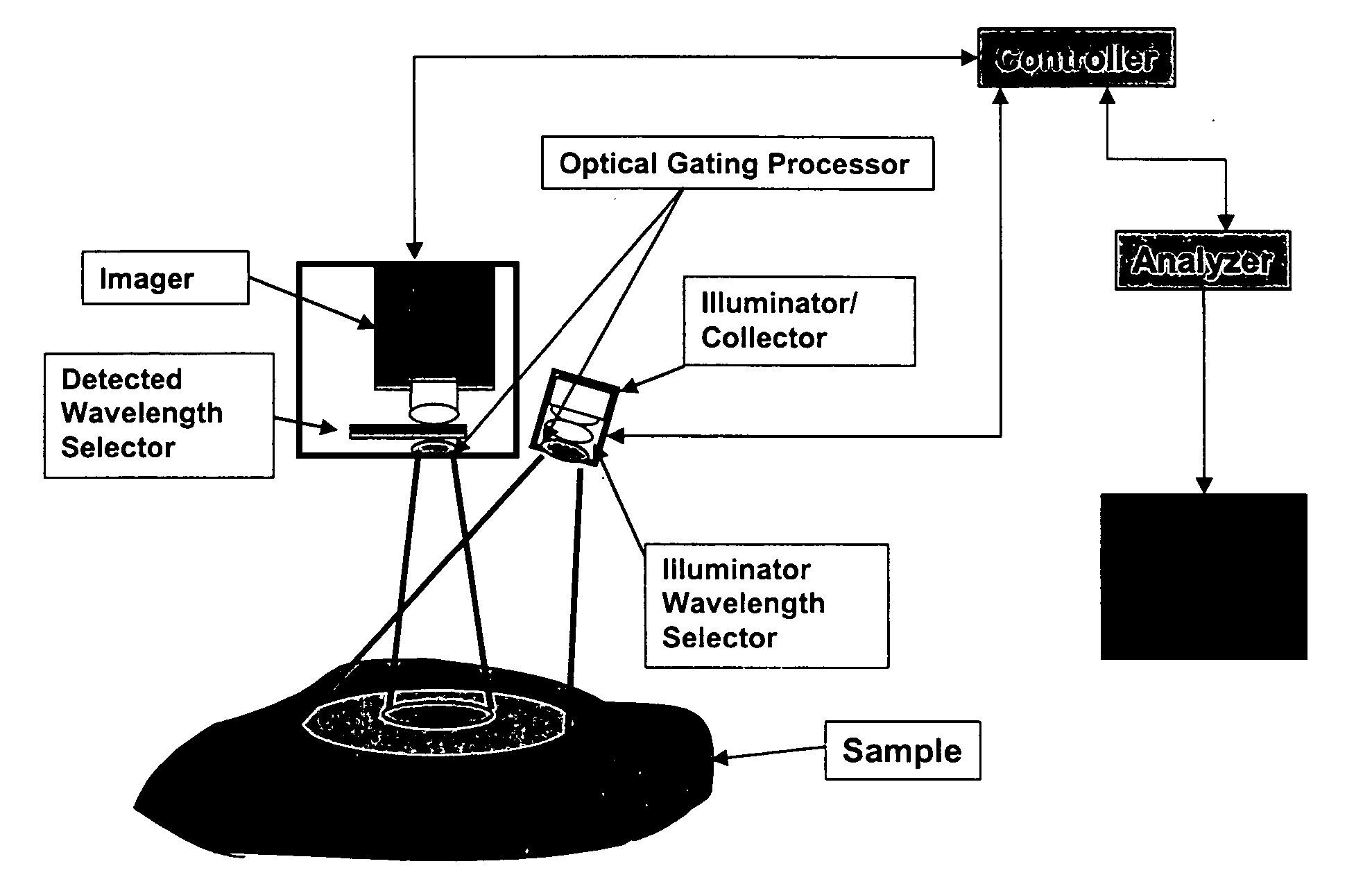
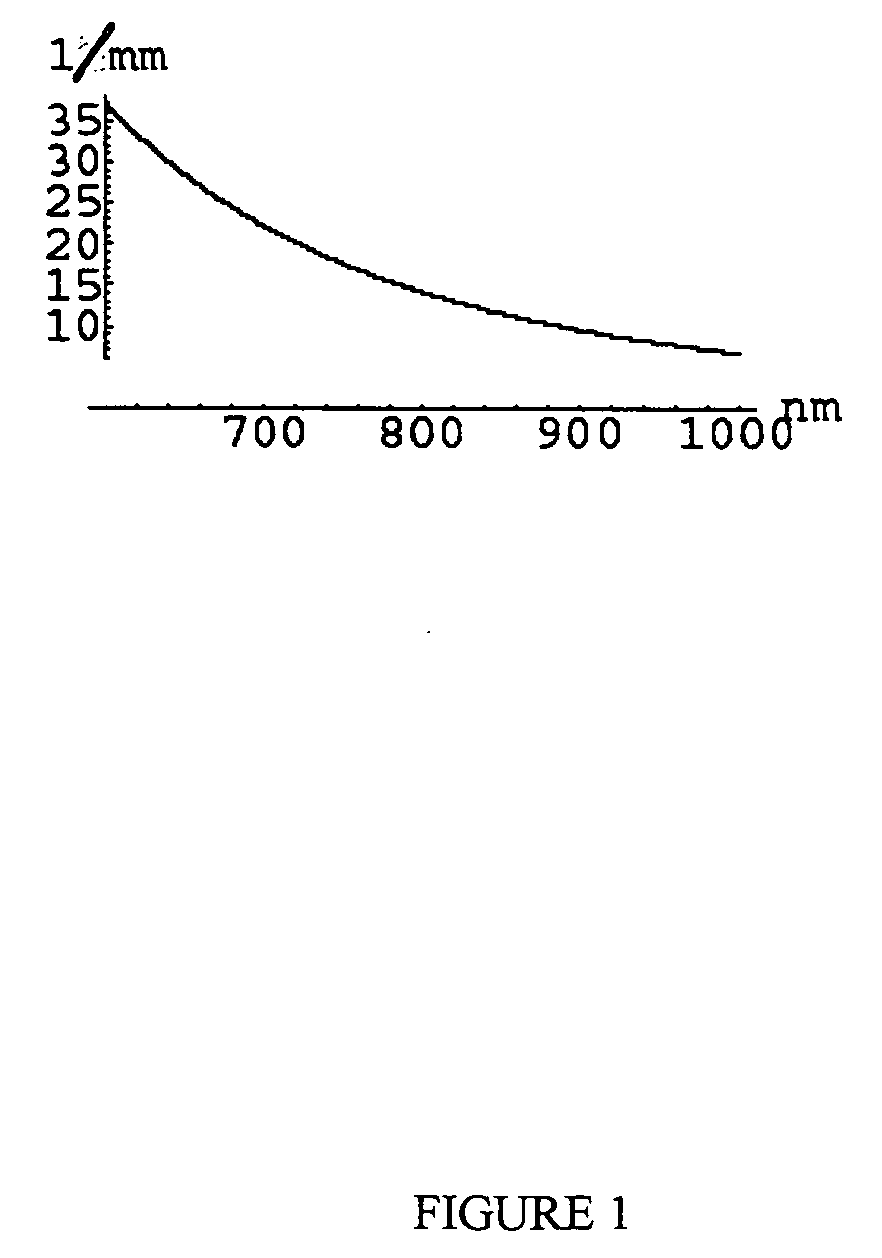
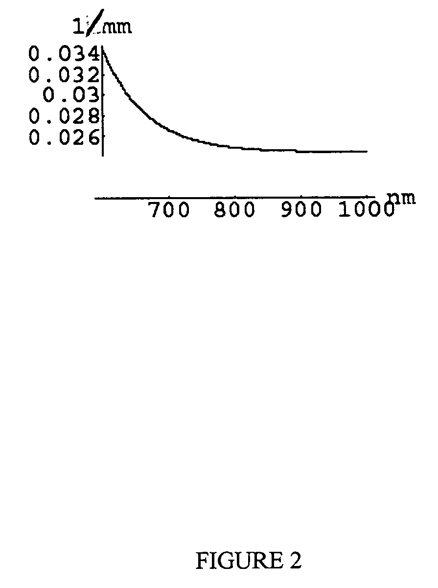
[0230] The disclosed invention may be used to obtain medically relevant functional and structural contrast from multispectral images. There are some commercially available multispectral imaging system and a number of ways of building multi-spectral imaging systems that are known to those skilled in the art. Typically such systems have a broad-band illumination source, a wavelength selecting device such as a filter wheel or tunable LCD filter on either the camera or illumination source, and an imager such as a CCD camera. The method was tested on data obtained using a CCD-based spectral imager that captures 512×512 pixel images at six wavelengths using a filter wheel on the camera (NIR at 700, 750, 800, 850, 850, 900 nm, with 50 nm FWHM and 1000 nm with 10 nm FWHM). While this is not generally the optimal choice of wavelengths and FWHM for the filters, it was sufficiently close to the optimal choice to show contrast as desired. The images have 16 bits per pixel...

example 2

CMOS Extended Dynamic Range

[0237] In biological tissue simulating phantom work that has already been completed, a commercially available CMOS-based color camera (Canon D-30) has been used to analyze the intensity-linearity of the pixels and the spectral response functions of the color filters on the pixels. [Additionally, a method was devised to access the raw 12 bit values, thereby bypassing the spatial filtering employed by the camera manufacturer to obtain satisfying color rendering] The method requires access to the raw pixel values (e.g., 12 bit on the Canon D-30) on the camera because it is necessary to bypass the spatial filtering which is often employed by consumer camera manufacturers to obtain satisfying color rendering. The method from obtaining raw values from an imaging device such as a CMOS device are described by the CMOS chip manufacturer's product data sheet or application instructions. Alternatively, some imaging systems include a method or instructions for obtain...


PUM


Abstract

A method for the evaluation of target media parameters in the visible and near infrared is disclosed. The apparatus comprises a light source, an illuminator/collector, optional illumination wavelength selector, an optional light gating processor, an imager, detected wavelength selector, controller, analyzer and a display unit. The apparatus illuminates an in situ sample of the target media in the visible through near infrared spectral region using multiple wavelengths and gated light. The sample absorbs some of the light while a large portion of the light is diffusely scattered within the sample. Scattering disperses the light in all directions. A fraction of the deeply penetrating scattered light exits the sample and may be detected in an imaging fashion using wavelength selection and an optical imaging system. The method extends the dynamic range of the optical imager by extracting additional information from the detected light that is used to provide reconstructed contrast of smaller concentrations of chromophores. The light detected from tissue contains unique spectral information related to various components of the tissue. Using a reiterative calibration method, the acquired spectra and images are analyzed and displayed in near real time in such a manner as to characterize functional and structural information of the target tissue.

Description

BACKGROUND OF THE INVENTION [0001] 1. Field of the Invention [0002] The field of the invention relates to quantitative optical spectroscopic imaging, and more particularly, to a method of obtaining functional and structural information from multispectral images of in situ analyzed multi-layered, optically dense samples. [0003] 2. Background Information [0004] Reflectance Spectroscopy [0005] In general, reflectance spectroscopy is concerned with the identification of a chemical structure comprising a sample through the use of reflected information. Diffuse reflectance spectroscopy is also generally known, and is widely used in the visible to infrared regions of the light spectrum to study materials to include, but not limited to, grains and other food products. [0006] In broad terms, diffuse reflectance spectroscopy utilizes the fact that the sample materials will tend to scatter light in a more or less random fashion. A fraction of the light will eventually be scattered back from th...

Claims


Application Information

Patent Timeline
IPC IPC(8): A61B5/00A61B6/00G01N21/47
CPCA61B5/0059G01N21/4738G01N21/4795A61B5/0075H04N5/335H04N25/00
Inventor HATTERY, DAVIDHATTERY, BRENDA
Owner APOGEE BIODIMENSIONS
Why Eureka
Try Eureka