Method for the automatic scaling verification of an image, in particular a patient image

a scaling verification and image technology, applied in image enhancement, instruments, medical/anatomical pattern recognition, etc., can solve the problems of change in imaging facilities, and typing errors during manual entry of calibration information, so as to increase the error reliability of the method preferably

Inactive Publication Date: 2005-12-22
SIEMENS AG
View PDF1 Cites 4 Cited by
  • Summary
  • Abstract
  • Description
  • Claims
  • Application Information

AI Technical Summary

Benefits of technology

[0022] The error reliability of the method preferably is increased by selecting a number of image regions at different positions inside the image range of the image, at least one shape segment respectively being selected inside each image region. This ensures that an erroneous evaluation quantity cannot be produced owing to a local individual anatomical difference of the patient's body, for example an abnormal bone growth in the region of a vertebra.

Problems solved by technology

Nevertheless, erroneous calibration information cannot entirely be ruled out.
Typical sources of error are, in particular, typing mistakes during the manual entry of calibration information, a change in the imaging facility without appropriate recalibration being carried out, or intentional or inadvertent data manipulation.
If a distance inside a patient image is determined incorrectly because of erroneous calibration, this can have critical consequences for the patient's health or life, especially when an incorrect recommendation to operate is made on the basis of the erroneously determined distance.

Method used


Image

  • Method for the automatic scaling verification of an image, in particular a patient image
  • Method for the automatic scaling verification of an image, in particular a patient image
  • Method for the automatic scaling verification of an image, in particular a patient image

Examples


Embodiment Construction

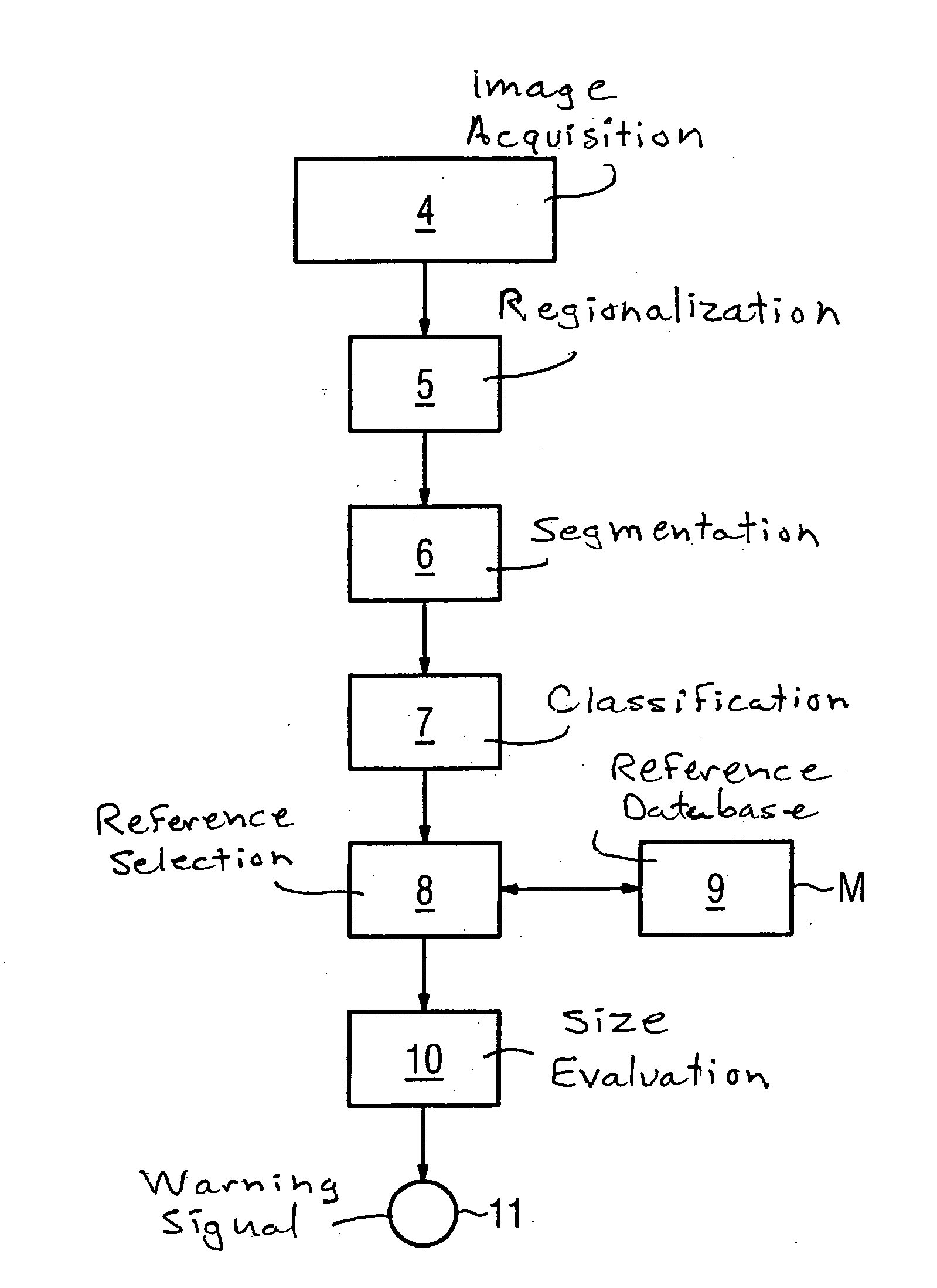
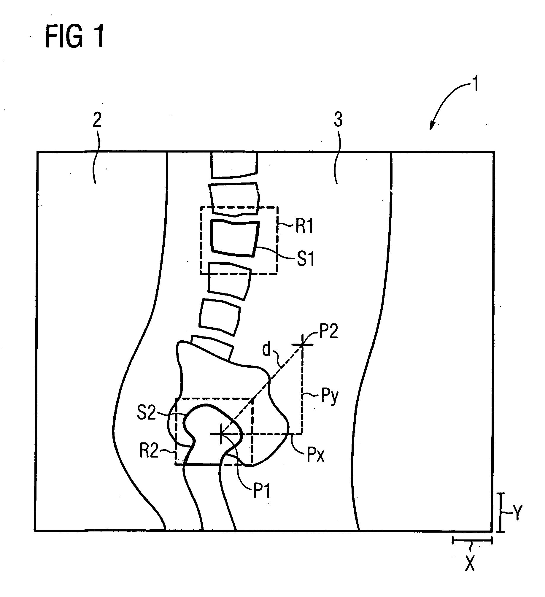
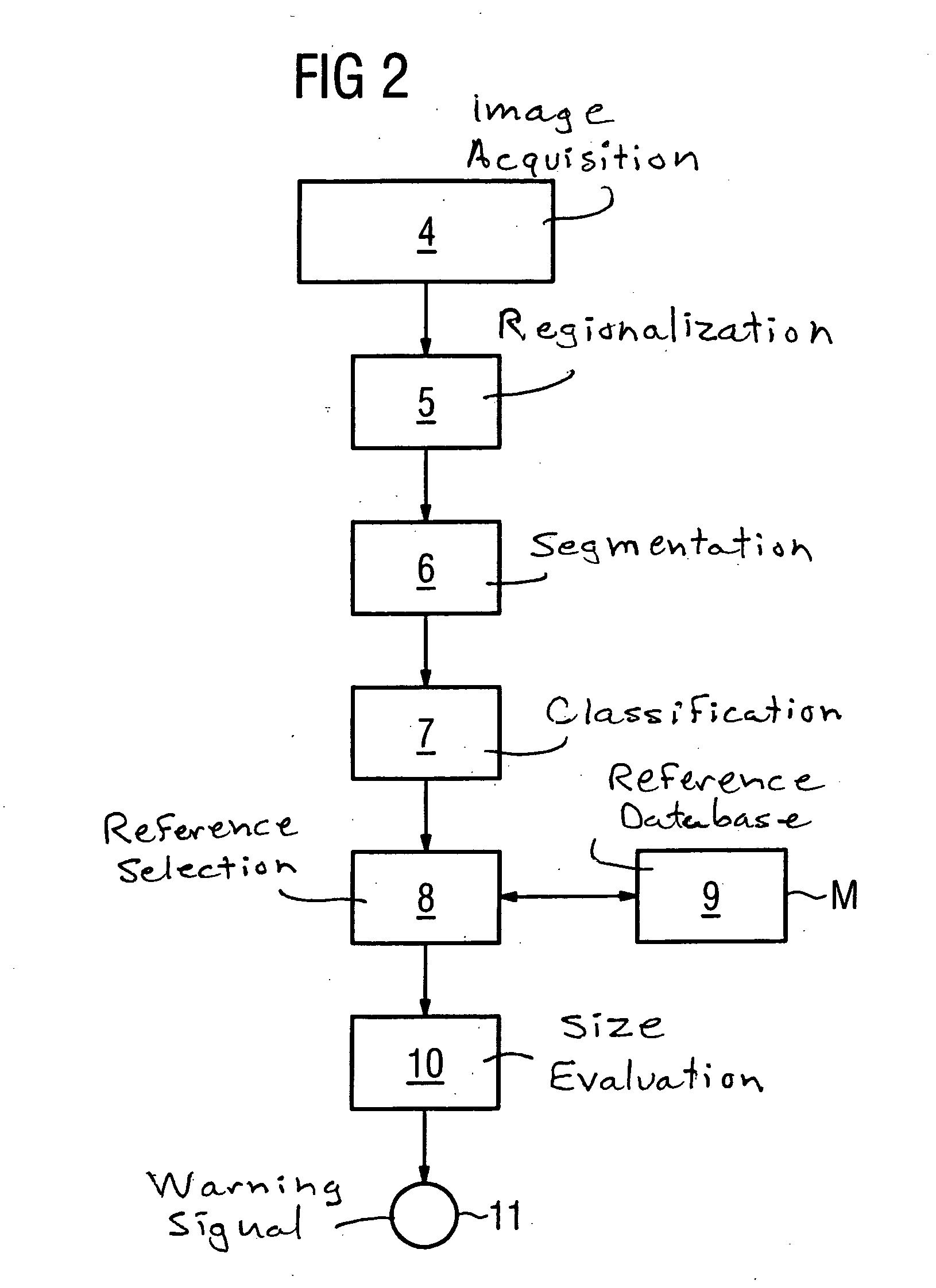
[0027] In order to illustrate the embodiment of the method respectively represented as simplified flowcharts in FIGS. 2 and 3, FIG. 1 schematically represents a two-dimensional patient image 1 as produced, for example, by a digital X-ray device. Such a digital patient image 1 includes a multiplicity of image points or pixels (not shown in detail) spatially arranged next to one another in a grid, each of which contains a color value or brightness value. The area covered by the image points (or the volume covered by the image points in the case of a three-dimensional patient image) is referred to as the image range 2.

[0028] The patient image 1 as represented shows a patient's body region 3 (in the example represented, the hip region in lateral projection).

[0029] The patient image 1 is assigned a horizontal scaling parameter X and a vertical scaling parameter Y. Each scaling parameter X, Y indicates the imaging scale of the patient image 1 in the corresponding space direction, in uni...


PUM

PropertyMeasurementUnit
medical imaging examinationaaaaaaaaaa
sizeaaaaaaaaaa
heightaaaaaaaaaa

Abstract

In a method to reliably avoid erroneous length determination in the analysis of an image, in particular a digital patient image formed in the course of a medical imaging examination method, by automatic scaling or by the scaling verification of such an image, a shape segment inside the image range of the image is identified and selected by electronic image processing, at least one classification parameter determined according to predetermined criteria is assigned to the shape segment, at least one reference segment comparable to the classification parameter or parameters is selected from a reference database, the size of the shape segment is evaluated using the selected reference segment, and an evaluation quantity characterizing the result of this evaluation is formed.

Description

BACKGROUND OF THE INVENTION [0001] 1. Field of the Invention [0002] The present invention relates to a method for the automatic scaling or for the scaling verification of an image, in particular a patient image formed in the course of a medical imaging examination. [0003] 2. Description of the Prior Art [0004] Digital medical imaging examination methods are gaining constantly increasing importance in medicine. Medical imaging examination methods” encompass X-ray examination methods, examination methods based on nuclear magnetic resonance, ultrasound examination methods and photographic examination methods (for example endoscopy). These include both examination methods that produce static images and those which produce dynamic, i.e. moving images. Imaging examination methods that produce three-dimensional patient images are also well established (for example computed tomography and magnetic resonance tomography). [0005] Image-assisted examination methods with similar requirements are...

Claims


Application Information

Patent Timeline
Patent Type & Authority Applications(United States)
IPC IPC(8): A61B5/107A61B6/02G01N23/04G06T3/40G06V10/32
CPCA61B5/1075A61B5/4504A61B5/7264G06T2207/30004G06K9/42G06K2209/05G06T3/0056G01N23/04G16H50/20G06V10/32G06V2201/03
Inventor BOTH, CARLOMUKKE, NORBERTRACZYNSKI, ARTUR
Owner SIEMENS AG
Why Eureka
Try Eureka