Inhibitors of macrophage migration inhibitory factor and methods for identifying the same

a macrophage migration and inhibitory factor technology, applied in the field of inhibitors of macrophage migration inhibitory factor and methods for identifying the same, can solve the problems of limited ability to evaluate the inhibition of mif to its cell surface receptor, significant toxicity, and inability to identify inhibitors that act intracellularly

Inactive Publication Date: 2005-12-29
AVANIR PHARMA
View PDF34 Cites 13 Cited by
  • Summary
  • Abstract
  • Description
  • Claims
  • Application Information

AI Technical Summary

Benefits of technology

[0007] As MIF has been identified in a variety of tissues and has been associated with numerous pathological events, there exists a need in the art to identify inhibitors of MIF. There is also a need for pharmaceutical compositions containing such inhibitors, as well as methods relating to the use thereof to treat, for example, immune related disorders or other MIF induced pathological events, such as tumor associated angiogenesis. The preferred embodiments may fulfill these needs, and provide other advantages as well.
[0033] In a seventeenth embodiment, a method is provided for suppressing an immune response in a warm-blooded animal, including administering to the animal an effective amount of the compound of the first embodiment.

Problems solved by technology

Hence, when its activities / gene expression are overinduced by the administration of excess exogenous glucocorticoids (for example when clinical indicated to suppress inflammation, immunity and the like), there is significant toxicity because MIF itself exacerbates the inflammatory / immune response.
Further, it remains unclear whether this enzymatic activity mediates the biological response to MIF and the activities of this protein in vitro and in vivo.
The ability to evaluate the inhibition of MIF to its cell surface receptor is also limited since no high affinity receptor is currently known.
Furthermore, inhibitors that act intracellularly are often toxic by virtue of their action on related targets or the activities of the target inside cells.
Small molecule inhibitors of the ligand receptor complex are difficult to identify let alone optimize and develop.

Method used


Image

  • Inhibitors of macrophage migration inhibitory factor and methods for identifying the same
  • Inhibitors of macrophage migration inhibitory factor and methods for identifying the same
  • Inhibitors of macrophage migration inhibitory factor and methods for identifying the same

Examples


example 1

[0199]

[0200] A solution of ethyl nitroacetate (6.70 ml, 60.1 mmol) in N,N-dimethylacetamide (35 ml) was treated with 95% NaH (1.52 g, 60.2 mmol) in portions. After evolution of hydrogen ceased, the reaction mixture was heated at 80° C. for 15 minutes. A solution of N-methylisatoic anhydride 1 (11.1 g, 62.5 mmol) in N,N-dimethylacetamide (65 ml) was added over a period of 15 minutes after which the reaction was heated at 120° C. for 18 hours. The solvent was removed by distillation, the residue dissolved in water, and acidified with 6 N HCl. The ensuing precipitate was collected and washed with water. Recrystallization of the remaining residue from CH2Cl2 / ether gave quinolinone 2a (5.75 g, 43%) as a yellow solid.

[0201] A solution of diethyl malonate (18.8 ml, 111 mmol) in N,N-dimethylacetamide (35 ml) was treated with 95% NaH (2.80 g, 111 mmol) in portions. After evolution of hydrogen ceased, the reaction mixture was heated at 80° C. for 15 minutes. A solution of N-methylisatoic an...

example 2

[0210] Macrophage Migration Assay

[0211] Macrophage migration is measured by using the agarose droplet assay and capillary method as described by Harrington and Stastny et al., J. Immunol. 110(3):752-759, 1973. Briefly, macrophage-containing samples are added to hematocrit tubes, 75 mm long with a 1.2 mm inner diameter. The tubes are heat sealed and centrifuged at 100×G for 3 minutes, cut at the cell-fluid interface and imbedded in a drop of silicone grease in Sykes-Moore culture chambers. The culture chambers contain either a control protein (BSA) or samples. Migration areas are determined after 24 and 48 hours of incubation at 37° C. by tracing a projected image of the macrophage fans and measuring the areas of the migration by planimetry.

[0212] Alternatively, each well of a 96-well plate is pre-coated with one microliter of liquid 0.8% (w / v) Sea Plaque Agarose in water dispensed onto the middle of each well. The plate is then warmed gently on a light box until the agarose drops ...

example 3

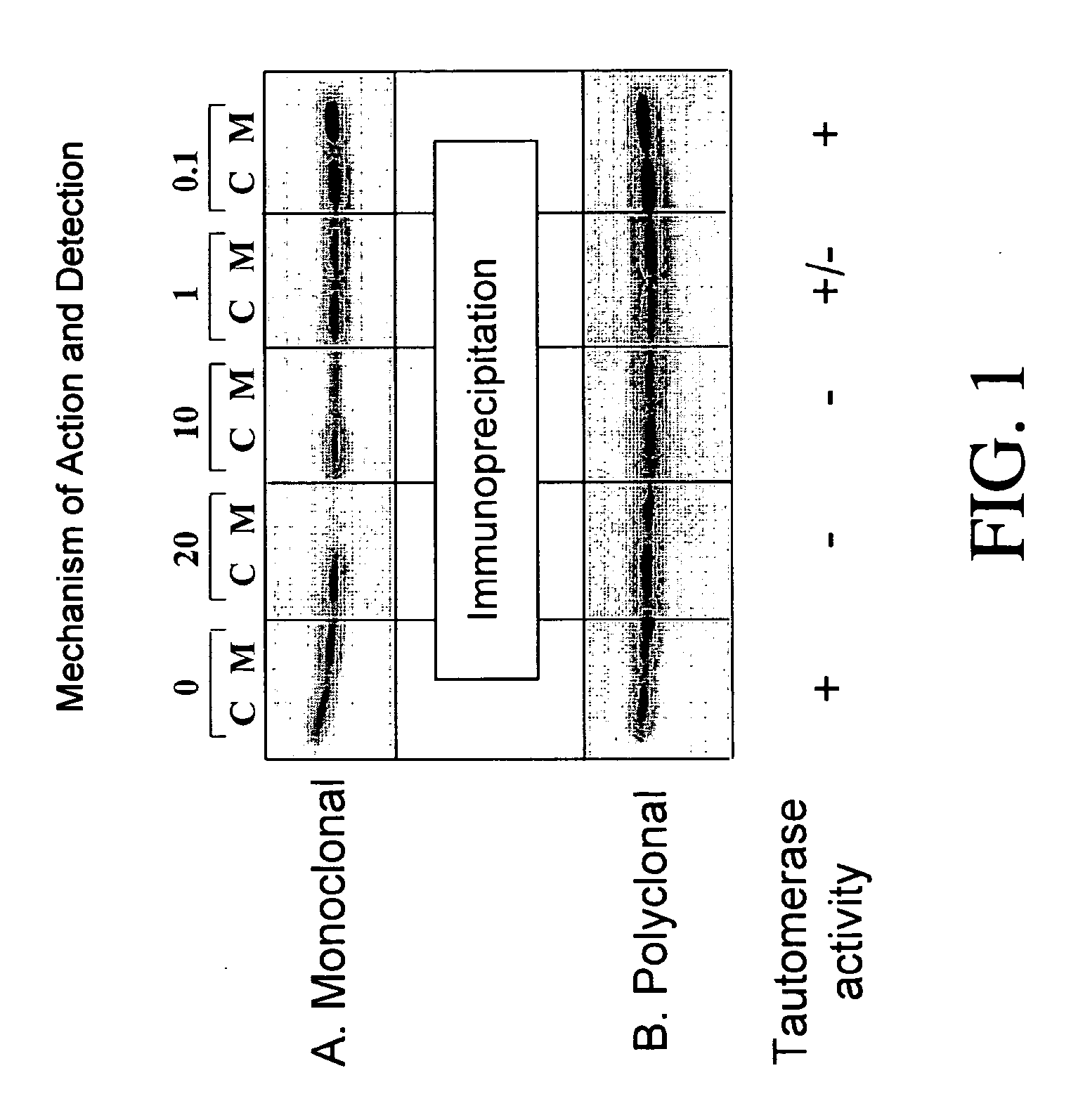
[0215] Tautomerase Assay

[0216] The tautomerization reaction is carried out essentially as described by Rosengren et al., Mol. Med. 2(1):143-149, 1996. D-dopachrome conversion to 5,6-dihydroxyindole-2-carboxylic acid is assessed. 1 ml sample cuvettes containing 0.42 mM substrate and 1.4 μg of MIF in a sample solution containing 0.1 mM EDTA and 10 mM sodium phosphate buffer, pH 6.0 are prepared and the rate of decrease in iminochrome absorbance is followed at 475 nm. L-dopachrome is employed as a control. In addition, the reaction products can be followed using an HPLC, utilizing a mobile phase including 20 mM KH2PO4 buffer (pH 4.0) and 15% methanol with a flow rate of 1.2 ml / min. Fluorimetric detection is followed at 295 / 345 nm.

[0217] Alternatively, the tautomerization reaction utilizing phenylpyruvate or (p-hydroxyphenyl)pyruvate is carried out essentially as described by Johnson et al., Biochem. 38:16024-16033, 1999. In this version, ketonization of phenylpyruvate is monitored at...


PUM

PropertyMeasurementUnit
molecular massaaaaaaaaaa
timeaaaaaaaaaa
timeaaaaaaaaaa

Abstract

Inhibitors of MIF are provided which have utility in the treatment of a variety of disorders, including the treatment of pathological conditions associated with MIF activity. The inhibitors of MIF have the following structures: including stereoisomers, prodrugs and pharmaceutically acceptable salts thereof, wherein n, R1, R2, R3, R4, X, Y and Z are as defined herein. Compositions containing an inhibitor of MIF in combination with a pharmaceutically acceptable carrier are also provided, as well as methods for use of the same.

Description

RELATED APPLICATION [0001] This application is a division of U.S. application Ser. No. 10 / 156,650, filed May 24, 2002, which claims the benefit of U.S. Provisional Application No. 60 / 293,642, filed May 24, 2001. All above-referenced prior applications are incorporated by reference herein in their entirety and are hereby made a portion of this specification.[0002] This invention relates generally to inhibitors of macrophage migration inhibitory factor (MIF), methods for identifying MIF inhibitors, and to methods of treating MIF-related disorders by administration of such inhibitors. BACKGROUND OF THE INVENTION [0003] The lymphokine, macrophage migration inhibitory factor (MIF), has been identified as a mediator of the function of macrophages in host defense and its expression correlates with delayed hypersensitivity, immunoregulation, inflammation, and cellular immunity. See Metz and Bucala, Adv. Immunol. 66:197-223, 1997. Macrophage migration inhibitory factors (MIFs), which are bet...

Claims


Application Information

Patent Timeline
Patent Type & Authority Applications(United States)
IPC IPC(8): A61K31/47A61K31/496A61K31/497C12Q1/02A61K45/00A61P1/04A61P3/10A61P5/08A61P5/46A61P9/00A61P11/00A61P11/06A61P13/12A61P19/02A61P25/28A61P29/00A61P35/00A61P37/06A61P43/00C07D215/42C07D215/46C07D215/54C07D401/12C07D405/12C07D409/12C07D409/14C07D413/12C07D417/12
CPCC07D215/42C07D215/54C07D401/12C07D405/12C07D409/12A61P1/04A61P11/00A61P11/06A61P13/12A61P19/02A61P25/28A61P29/00A61P35/00A61P37/06A61P43/00A61P5/08A61P5/46A61P9/00A61P3/10C07D401/04
Inventor GAETA, FEDERICOBAIRD, ANDREWANCHIN, JERRYYING, WENBINFLORKIEWICZ, ROBERTSIRCAR, JAGADISHK.C., SUNIL
Owner AVANIR PHARMA
Why Eureka
Try Eureka